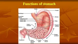
Various Stomach Anatomy Slides of Different Animal Species
Explore a series of detailed histological slides showcasing the stomach anatomy of different animal species including cats, rabbits, dogs, pigs, sheep, and goats. The images highlight key features such as gastric pits, mucosal layers, chief and parietal cells, proper gastric glands, cardiac and pyloric glands, mucus neck cells, and different regions of the stomach with distinct cellular compositions.

Download Presentation

Please find below an Image/Link to download the presentation.
The content on the website is provided AS IS for your information and personal use only. It may not be sold, licensed, or shared on other websites without obtaining consent from the author. Download presentation by click this link. If you encounter any issues during the download, it is possible that the publisher has removed the file from their server.
E N D
Presentation Transcript
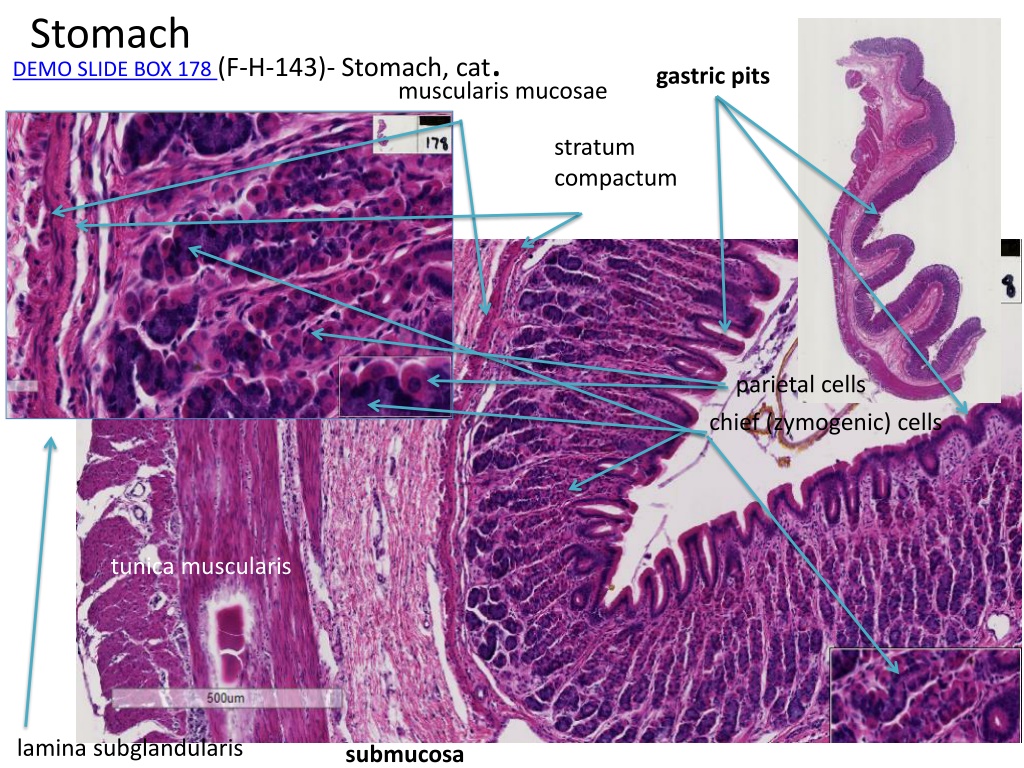
Stomach DEMO SLIDE BOX 178 (F-H-143)- Stomach, cat. gastric pits muscularis mucosae stratum compactum lamina propria parietal cells chief (zymogenic) cells tunica muscularis lamina subglandularis submucosa
DEMO SLIDE BOX 178 (F-H-143)- Stomach, cat. CIRCULAR, LONGITUDINAL, CIRCULAR muscularis mucosae (three layers) stratum compactum Muscularis mucosae muscularis mucosae
DEMO SLIDE BOX 178 (F-H-143)- Stomach, cat. Not well defined stratum compactum muscularis mucosae (three layers) chief (zymogenic) cells CIRCULAR, LONGITUDINAL, CIRCULAR parietal cells
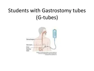
Mucus neck cells Fundic stomach, rabbit (toluidine blue) 244 Enteroendocrine cells Chief cells Parietal cell
DEMO SLIDE BOX 51 - Stomach, dog. pyloric gland region. pyloric glands =MUCOUS Gastric pits thickened tunica muscularis = PYLORIC SPHINCTER solitary lymph nodules
DEMO SLIDE BOX 51 - Stomach, dog. PROPER GASTRIC GLANDS HAVE PARIETAL AND CHIEF CELLS PRESENT PROPER GASTRIC GLANDS with PARIETAL AND CHIEF CELLS PRESENT
DEMO SLIDE BOX 163 (P001-H-141,142,144) - Stomach, pig. PROPER PROPER: PARIETAL AND CHIEF CELLS
DEMO SLIDE BOX 163 (P001-H-141,142,144) - Stomach, pig. CARDIAC: GLANDS HAVE CELLS WITH ROUND NUCLEI CARDIAC PYLORIC PROPER PYLORIC: GLANDS HAVE CELLS WITH FLAT NUCLEI the serous-type cells lining in pits and high density of lymphocytes
DEMO SLIDE BOX 164 (O-001-H-142AB) - Abomasum, sheep. spiral folds. PROPER
Slide #179 (GT-1 174)- Abomasum, goat. abomasum not taken from the proper gastric gland region as no chief or partial cells serosa Smooth muscle Of externa
Slide #179 (GT-1 174)- Abomasum, goat. Three layers of tunica muscularis Nerve cell body In submucosa nerve Nerve cell body in tunica muscularis capillary lymphatic venule arteriole muscularis mucosae
Slide # 29. Esophagus, proventriculus, and gizzard, chicken thick tunica muscularis SMOOTH MUSCLE gizzard = ventriculus stratified squamous epithelium simple columnar epithelium The proventricular glands in the lamina propria are the outstanding features of this organ. KOILIN
DEMO SLIDE 177 (831) - Esophagus, proventriculus, and gizzard, chicken gizzard Esophagus proventriculus
DEMO SLIDE BOX 53 - Demo slide # 53a Proventriculus, chicken. lymphatic nodules Side of the gizzard / ventriculus submucosa layer is very thin
DEMO SLIDE BOX 54 - Demo slide # 54a. Gizzard, chicken. gizzard = ventriculus tunica muscularis is very thick and has a diamond appearance simple tubular glands lined with simple cuboidal epithelium Submucosa diamond appearance of tunica muscularis CCT thick koilin, secreted by the glands of the lamina propria. muscularis mucosae is absent
DEMO SLIDE BOX 55 Demo slide #55. Gizzard, chick. muscularis mucosae is absent CCT is stained green (blue) tunica muscularis diamond appearance created by the CCT interlacing the tunica muscularis not seen in developing gizzard better seen in Demo slide 54a below
Slide #40 (CK1 75/98/115B) Proventriculus, chicken. Gizzard Esophagus Proventriculus thick koilin, secreted by the glands
Demo slide # 53b- Gizzard???, chicken Proventriculus
Demo slide 54a thick koilin tunica muscularis diamond appearance created by the CCT interlacing the tunica muscularis not seen in developing gizzard
Slide #92 (Sp 804-102F). Rumen, sheep. Ruminant Forestomach ruminal papilla tunica muscular is arranged in two distinct layers keratinized stratified squamous epithelium mesothelium No muscularis mucosae is present serosa submucosa
DEMO SLIDE BOX 52 - Demo slide # 52. Rumen, cow. papillae The stratified squamous epithelium varies in thickness and the keratin is not readily seen serosa tunica muscularis is composed of two layers of smooth muscle numerous papillae
DEMO Slide #228 (O-001-H-142RU). Rumen, sheep. keratinized stratified squamous epithelium numerous papillae
Slide #90 (1124). Reticulum, steer. large projections are termed reticular crests, while the smaller ones are papillae. muscularis mucosae of the reticulum is incomplete no muscularis mucosae is present in the papillae.
Slide #32 (O-001-H-142-RE). Reticulum, sheep. no muscularis mucosae is present in the shorter papillae. muscularis mucosae muscularis mucosae of the reticulum is incomplete in the reticular crests
Slide #195 (BV 5 101b). Reticulum, cow. muscularis mucosae of the reticulum is incomplete in the reticular crests mesothelium serosa
Slide #191 (GT-1-93). Omasum, goat. mucosal projections of this compartment are quite long ( spider leggy ) and are termed omasal laminae muscularis mucosae is complete and strands of smooth muscle derived from the tunica muscularis are also present in the laminae.
Slide #196 (BV 5 88a). Omasum, cow. serosa tunica muscularis large muscular artery muscularis mucosae is complete nerve omasal laminae CCT unilocular adipose cells
Slide #91 (98-B-OM-8A)- Omasum, cow. muscularis mucosae is complete and strands of smooth muscle derived from the tunica muscularis are also present in the laminae.
Slide #33 (O-001-H-142-RE) - Omasum, sheep. muscularis mucosae is complete omasal laminae

- Stomach, cat.")
- Stomach, cat.")
")


 -")
 -")
 - Abomasum,")
- Abomasum, goat.")
- Abomasum, goat.")

 - Esophagus, proventriculus,")


 Proventriculus, chicken.")


. Rumen, sheep.")

. Rumen, sheep.")
. Reticulum, steer.")
. Reticulum, sheep.")
. Reticulum, cow.")
. Omasum, goat.")
. Omasum, cow.")
- Omasum, cow.")
 - Omasum, sheep.")