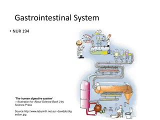
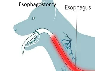
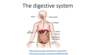
Radiology Imaging of Esophagus and Stomach by Dr. A. Alhawas
View detailed radiology imaging of the esophagus and stomach conducted by Dr. A. Alhawas, showcasing various anatomical structures such as the splenic artery, abdominal aorta, common hepatic artery, and more. Explore pathologically changed layers in pyloric stenosis, arterial blood supply to the pylorus and first part of the duodenum, and structures forming the stomach bed.

Download Presentation

Please find below an Image/Link to download the presentation.
The content on the website is provided AS IS for your information and personal use only. It may not be sold, licensed, or shared on other websites without obtaining consent from the author. Download presentation by click this link. If you encounter any issues during the download, it is possible that the publisher has removed the file from their server.
E N D
Presentation Transcript
Imaging of esophagus and stomach Radiology department Dr. A. Alhawas
3 1 2 4 1. Splenic artery 2. Abdominal aorta 3. Common hepatic artery 4. Gastro-duodenal artery
1. 2. 3. 4. 5. Abdominal aorta. Common hepatic artery. Splenic artery. Gasto-duodenal artery. Left gasto-epiploic artery.
1 4 5 2 6 3 1. 2. 3. 4. 5. 6. Left main pulmonary artery. Esophagus. Thoracic arota. Right main pulmonary artery. Left main bronchus. Azygous vien.
1 3 4 2 5 1. 2. 3. 4. 5. Stomach Splenic artery / short gastric artery. Gastro-esophageal junction. Inferior vena cava. Aorta.
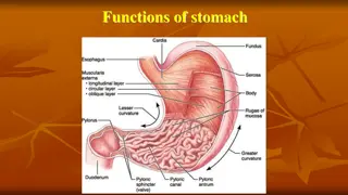
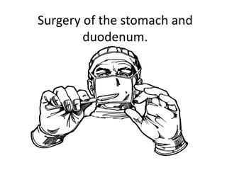
State which layers are pathologically changed in pyloric stenosis. both circular and longitudinal layers . State the name of arteries supplying the pylorus and the first part of the duodenum. State four (4) structures forming the stomach bed: 1. Pancreas ( body and tail ). 2. Spleen. 3. Left kidney. 4. Left adrenal gland. 5. Left crus of the diaphragm. 6. Splenic artery 7. Transverse mesocolon.