Anatomy and Blood Supply of the Equine Stomach
The equine stomach is relatively small compared to the horse's body size, with distinct regions such as the cardia, fundus, body, and pyloric region. It is located on the left side of the abdomen, under the ribs. The stomach's blood supply includes branches from the aorta, splenic artery, and hepatic artery, ensuring proper circulation. Understanding the anatomy and blood flow of the equine stomach is essential for the overall health and function of horses.
- Equine Stomach Anatomy
- Horse Digestive System
- Stomach Blood Supply
- Equine Health
- Veterinary Medicine

Download Presentation

Please find below an Image/Link to download the presentation.
The content on the website is provided AS IS for your information and personal use only. It may not be sold, licensed, or shared on other websites without obtaining consent from the author. Download presentation by click this link. If you encounter any issues during the download, it is possible that the publisher has removed the file from their server.
E N D
Presentation Transcript
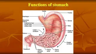
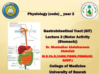
Anatomy The equine stomach is small relative to the body size of the horse, having a capacity of approximately 5 to 15 It is located on the left side of the abdomen under the cover of the ribs, with only the pyloric region of the stomach to the right side of the midline. Its most caudal component is the fundus, which lies adjacent to the 14thand 15th rib spaces The stomach can be divided into 1- cardia at the opening of the esophagus, 2- fundus (which forms a blind sac), 3- body, 4- pyloric region
Anatomy . The stomach is sharply curved at its lesser curvature so that the cardia and pyloric regions lie adjacent to each other. The cardia is attached to the diaphragm by the gastrophrenic ligament. This ligament is a continuation of the phrenicosplenic ligament and the gastrosplenic ligament on the left side of the abdomen. The greater omentum(omental bursa) attaches along the greater curvature of the stomach, and it blends into the gastrophrenic ligament. The epiploic foramen is also bordered dorsally and ventrally by the caudal vena cava and portal vein, respectively. The lesser omentum, which connects the stomach and duodenum to the liver, consists of the hepatogastric and epatoduodenal ligaments.
Blood supply Aorta -- celiac artery 1-splenic, 2-hepatic, 3- left gastric arteries Splenic artery --tributaries left limb of the pancreas and spleen--left gastroepiploic artery, left gastroepiploic artery supplies the greater curvature of the stomach and anastomoses with the right gastroepiploic artery hepatic artery --branches to the liver and gallbladder --right gastric artery, which supplies blood to the pylorus and pyloric antrum, anastomoses with the left gastric artery along the lesser curvature
Blood supply The venous drainage of the stomach to the portal vein is through the gastrosplenic vein on the left and gastroduodenal vein on the right Lymphatic drainage of the stomach is through the gastric and splenic lymph nodes to the hepatic lymph nodes The stomach is innervated by parasympathetic fibers of the vagus nerves and sympathetic fibers of the celiac plexus
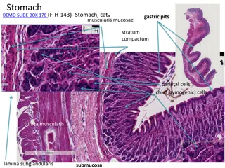
Gastric Layers The stomach wall 1-serosa, 2-muscle,3-submucosa, thin elastic layer of areolar tissue 4- mucosa. The muscular composition is divided into three layers 1- The longitudinal layer 2-The inner circular layer(gastroesophageal (cardiac)Sphincter) Grinding function of the antrum 3-The oblique muscle fibers
Diagnostic Techniques Endoscopy Before gastric endoscopy, foals up to 20 days of age should be held off feed for 3 hours, whereas older foals require up to 10 hours to sufficiently empty the stomach. Adult horses should be held off feed for 24 hours to allow complete visualization of the stomach. 3-m endoscope is required to adequately visualize the stomach, including the pylorus
Ultrasonography Ultrasonography can be used to image the wall of the stomach, and it may be particularly useful in foals with suspected gastric outflow obstruction. The stomach is best imaged from the left side of the abdomen between rib spaces 8 and 14. If gastric outflow obstruction is present, a distended stomach with a gas fluid interface may be detected.
Radiography Contrast radiography can be performed to allow visualization of the stomach. barium sulfate solution functional gastric emptying can be determined by the administration of barium sulfate after a 12- to 18-hour fasting period. In normal horses, barium appears in the small intestine by 10 minutes, and none remains within the stomach by 35 minutes. With pyloric outflow obstruction, barium may remain in the stomach for up to 8 hours. Barium sulfate solutions should be diluted 1 : 1 with water, and a volume of approximately 1 L should be administered into the thoracic portion of the esophagus by nasogastric tube. Double-contrast radiography with insufflation of air, followed by administration of a barium sulfate suspension, can enhance visualization of the stomach. This technique has been useful in the diagnosis of gastric outflow obstruction and gastric neoplasia.
Mechanism of ulcer injury of the gastric mucosa may be entirely different from those inducing injury to stratified squamous mucosa. the majority of gastric mucosal ulcers is induced by infection with Helicobacter pylori, which has the effect of raising gastric pH because of disruption of gastric glands. Such an infection also induces an inflammatory response that causes damage, In particular, H. pylori containing the cagA gene is most pathogenic. However, there is very little evidence that this organism is involved in gastric ulcers in domestic animals. , ulceration most likely develops from an imbalance between protective mechanisms and injurious factors, which include gastric acid, bile, and nonsteroidal anti-inflammatory drugs (NSAIDs)
Disorders Gastric Ulcer Clinical Syndromes The clinical syndromes of gastric ulceration are age dependent; the age categories being neonates, weanling foals, and horses older than 1 year. In neonates, ulceration of the glandular mucosa is the most clinically important. Clinical signs include poor appetite, colic, and diarrhea, but foals may have ulcers and not demonstrate overt clinical signs of gastric pain. The pH of neonatal gastric contents is not typically as low as that of older foals or adults, suggesting that other factors may be important in the pathogenesis of ulceration.
Treatment is aimed at elevating the pH of the gastric contents, which may be achieved with a number of histamine receptor (H2) antagonists, such as ranitidine (6.6 mg/kg, PO every 8 hours, or 1.5 to 2 mg/kg IV every 6 to 8 hours) or proton pump inhibitors such as omeprazole (2 to 4 mg/kg PO every 24 hours)
Gastric Impaction Impaction of the stomach typically consists of excessive dry, fibrous ingesta, but it may also consist of ingested materials that form a mass, such as persimmon seeds or mesquite beans. Other feeds that tend to swell after ingestion, such as wheat, barley, and sugar beet pulp, may also cause impaction. Furthermore, dental disease may increase the likelihood of gastric impaction because of improper chewing of feed. Clinical signs include colic that ranges from acute and severe to chronic and mild. The diagnosis is frequently made at the time of surgery, although endoscopy reveals gastric impaction and may provide information on the specific nature of the impaction.
Medical treatment includes nasogastric intubation and frequent attempts at softening the ingesta with water, followed by refluxing the fluid contents, or by using back-and-forth agitating movements of water with a 16-ounce dose syringe attached to the nasogastric tube. Nasogastric lavage with a carbonated cola soft drink was successfully used in a pony with persimmon seed impaction of the stomach Surgery, the impaction can be massaged and infused, most commonly via insertion of a needle adjacent to the greater curvature, followed by infusion of a balanced polyionic fluid such as saline. There is also a report including the details of a pony and a horse in which the impacted stomach was packed off from the abdomen with towels, and an incision was made parallel and caudal to the attachment of the omentum on the greater curvature of the stomach.
Chronic Gastric Impaction and Dilation Typically, gastric impaction develops relatively quickly and the diagnosis is often made at surgery. In contrast, chronic impaction and dilation of the stomach develops slowly over weeks or months with minimal clinical signs of abdominal pain. Clinical signs are often mild, with weight loss, reduced performance, bruxism, and salivation reported. Gastric endoscopy may reveal fibrous ingesta, which does not change after 24-hour starvation. A large mass may be palpable per rectum. Abdominal radiographs or ultrasonography may demonstrate a large distended and impacted stomach. Gastrotomy to remove ingesta and partial gastrectomy to remove flaccid stomach wall have been unsucessful.
Gastric Rupture Rupture of the stomach appears to have two general causes: primary, excessive accumulation of ingesta, secondary, in association with another causative condition, such as obstruction of the small intestine. The site of the rupture is most commonly the greater curvature, although other sites of rupture have been identified. The condition is almost universally fatal, because release of stomach contents into the abdomen through the gastric tear causes septic shock that cannot be adequately reversed with abdominal lavage and repair of the defect.
Gastric Neoplasia Neoplasia of the stomach is rare. The most common form of gastric neoplasia is squamous cell carcinoma, which typically forms in the cardia of the stomach. In one report, a tumor encircled the esophagus at the cardia of the stomach, where it caused recurrent esophageal obstruction. Other neoplasms that have been identified in the stomach include leiomyosarcoma (leiomyoma), mesothelioma, and adenocarcinoma. Clinical signs may include anorexia, weight loss, abdominal distention, abnormal chewing behavior, lethargy, coughing, hypersalivation, colic, dysphagia, fever, and ventral abdominal edema. The diagnosis can be based antemortem on results of gastric endoscopy and biopsy or on thoracoscopy and biopsy, In addition, approximately 50% of horses have had neoplastic cells evident in abdominal fluid or in thoracic fluid.
Gastric Outflow Obstruction The result of pyloric stenosis, which can be caused by congenital muscular hypertrophy, or by development of a mass at the pylorus that reduces gastric outflow. A mass may develop at the pylorus associated with gastroduodenal ulceration or neoplasia. Clinical signs include weight loss, reduced appetite, abdominal pain, teeth grinding, ptyalism, frequent recumbency, and poor performance. Foals with gastric outflow obstruction are typically 2 to 6 months of age. foals may also have a history of enteritis and an absence of clinical signs typical of foals with gastric ulcers. Fot diagnosed using endoscopy, radiography, ultrasonography, and gastric emptying tests, as described previously
Medical and surgery treatment Decompression of the stomach, antiulcer medications, broadspectrum antibiotics, prokinetics, and intravenous fluids. Surgery is indicated if medical treatment does not reverse clinical signs within a short period. The principle of surgery for treatment of gastric outflow obstruction is bypass of the pylorus, typically by performing a gastrojejunostomy. Pyloric stenosis has been relieved in a 2-month-old Thoroughbred by a modification of the Heineke-Mikulicz technique, in which a full-thickness longitudinal incision through the pylorus was closed transversely. The pylorus was mobilized by severing the hepatoduodenal ligament.
Alternatively, a series of bypass techniques can be performed depending on the location of the obstruction. bypass procedures included 1- gastroduodenostomy, 2- duodenojejunostomy, 3- gastrojejunostomy These anastomoses can be hand-sewn or performed with automated stapling equipment, hand-sewn anastomoses may be simpler to perform because of the limited space within a foal s abdomen, which reduces maneuverability of larger stapling instruments. Although a three-layer hand-sewn technique has been described (seromuscular, muscular, and mucosal layers),