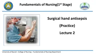
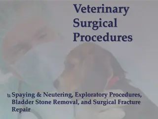
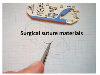
Esophagostomy Surgical Procedure Overview
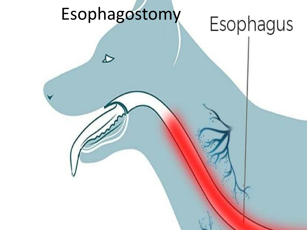
Esophagostomy is a surgical procedure mainly performed to address foreign bodies or esophageal diverticula in animals like horses, cattle, dogs, and cats. The operation involves accessing the esophagus, removing the foreign body, and suturing the esophageal wall. Key steps include making an incision in the jugular groove, identifying the esophagus by its color, and carefully suturing the mucosa and muscular layers. Blood vessels supplying the esophagus are also crucial in the procedure. Overall, esophagostomy is a delicate surgery that requires precise technique and attention to detail.

Download Presentation

Please find below an Image/Link to download the presentation.
The content on the website is provided AS IS for your information and personal use only. It may not be sold, licensed, or shared on other websites without obtaining consent from the author. Download presentation by click this link. If you encounter any issues during the download, it is possible that the publisher has removed the file from their server.
E N D
Presentation Transcript
Indication 1. foreign bodies (potatoes, turnips ect) in horses and cattle lodged in the cervical part of the esophagus .in dogs and cats bones are lodged in the cervical and thoracic parts of the esophagus. 2. esophageal diverticula's.
operation procedure the operation is performed from the left side of the neck or from the site of the foreign body in recumbent position under general anesthesia
blood vessels of the esophagus cervical portion: cranial and caudal thyroid arteries and esophageal branch of carotids thoracic portion: bronchesophageal artery ventral portion: left gastric artery
Surgical procedure the site of operation clipped , shaved , and disinfected 10-20 cm long incision should be made longitudinally in the jugular groove the skin incision should be made between the jugular vein and the edge of sternomandibular muscle far away from the carotid artery
Surgical procedure incision is continued through the cutaneous muscles and blunt dissection is used until reach to the lateral surface of the trachea the esophagus is recognized by stomach tube , it is pale reddish color
Surgical procedure the esophagus is opened longitudinally to allow the remove of the foreign body without tearing the wall ,the foreign body is removed by rough thumb forceps
Surgical procedure the wound of the esophageal wall is sutured as the following patterns A- mucosa is suture alone with cat gut 3-0 usp with a simple interrupted pattern and the knot should be inside the lumen. B- muscular layer of the esophagus is sutured with interrupted or continuous suture pattern with non absorbable suture material the muscles is sutured with simple continuous pattern with catgut
Surgical procedure the operation area should be flushed with solution containing broad spectrum antibiotics the skin is sutured by simple interrupted pattern
the structures you found in esophagostomy skin cutaneous muscles sternomandibular muscles jugular vein vagus nerve carotid artery recurrent nerve trachea esophagus
post operative care systemic antibiotics (penstrept): 10000-20000 IU/kg body weight for 3 days post operatively keep the animal on fluid therapy at least 4 days and then soft fluid are given to the animal and semi sold food in the thirds week and forth week
complications: constricting scar at the site of incision esophageal fistula hemorrhage infection


![Briefing on the Criminal Procedure Amendment Bill [B12-2021] to the Portfolio Committee on Justice and Correctional Services](/thumb/157093/briefing-on-the-criminal-procedure-amendment-bill-b12-2021-to-the-portfolio-committee-on-justice-and-correctional-services.jpg)