Radiological Investigation of Hepatobiliary System and Imaging Modalities Overview
Exploring the hepatobiliary system through radiology, this lecture covers the anatomy, modalities like X-ray, Ultrasound, CT scan, MRI, and nuclear scan used for imaging, their advantages, disadvantages, and indications. It delves into the significance of liver, gallbladder, and biliary duct imaging, providing insights into each modality's role in diagnostics.
Download Presentation
Please find below an Image/Link to download the presentation.
The content on the website is provided AS IS for your information and personal use only. It may not be sold, licensed, or shared on other websites without obtaining consent from the author. Download presentation by click this link. If you encounter any issues during the download, it is possible that the publisher has removed the file from their server.

Presentation Transcript
Radiology & investigation of Radiology & investigation of hepatobiliary hepatobiliary system system Dr.Reshaid Aljurayyan Assistant Professor department of radiology college of medicine King Saud university
Lecture outline: Lecture outline: What is the hepatobiliary system (HBS)? Radiological modalities used in imaging HBS. Advantages and Disadvantages of each radiology modality. Indications of imaging HBS.
What HBS incudes? What HBS incudes? It includes liver, gallbladder and biliary ducts.
What are What are Radiological modalities Radiological modalities used in imaging HBS ? imaging HBS ? used in X Ray. Ultrasound. Computed tomography CT scan. Magnetic resonance imaging MRI. Nuclear scan.
What are What are Radiological modalities Radiological modalities used in imaging HBS ? imaging HBS ? used in X Ray. Ultrasound. Computed tomography CT scan. Magnetic resonance imaging MRI. Nuclear scan. ALL modalities can be used
What is this? What is this?
What is this? What is this? Abdomen x-ray OR Abdomen radiography
What is this ???? What is this ????
X ray was first observed and documented in 1895 by Wilhelm Conrad Roentgen First x ray taken in history
What is X ray? What is X ray? It is energetic form of electromagnetic and ionizing radiation that can penetrate solid objects and used to take images of the human body.
X X- -Ray language: Ray language: Radio-lucent = black Radio-opaque= white
X X- -Ray: Ray: Advantages: Quick and widely available Cheap Can be done bedside (portable) Disadvantages: Use ionizing radiation Very poor in tissue details including HBS Very limited in detecting gallbladder stones
What is this? ULTRASOUND
What is US? What is US? A diagnostic technique in which high-frequency sound waves penetrate the body and produce multiple echo patterns. Diagnostic Medical applications in use since late 1950 s
Ultrasound Ultrasound Advantages: No radiation. Widely available. Relatively cheap. Very good in evaluating abdomen solid organs. Can be done bedside (portable). Disadvantages: Operator dependent. Very limited in evaluating structures with air ( e.g. bowel) or calcification (e.g. bone).
Echo patterns Hyper-echoic = White Hypo-echoic = Light Grey An-echoic = Black Acoustic shadow: black band behind dense object (e.g. stone)
B- MODE DUPLEX COLOR DOPPLER
B- MODE DUPLEX
What is this? What is this?
What is this? What is this? CT scan = Computed Tomography
What is CT scan? What is CT scan? A CT scan is computer-processing of many X-ray images taken from different angles to produce cross-sectional images. CT scan can be done with and without intravenous IV contrast. CT scan is limited in evaluating gallstones, Why?
What is different between the tow iamges? ? With IV contrast Without IV contrast
Computed tomography CT scan: Computed tomography CT scan: Advantages: Very good in evaluating solid organs. Available more then MRI. Disadvantages: Use ionizing radiation. Less available then x-ray and US. Relatively expansive. Intravenous contrast maybe harmful in patient with impaired renal function..
CT language CT language Hyper-dense = white Hypo-dense=black to grey
What is this? What is this?
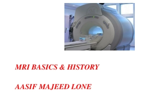
What is this? What is this? Magnetic resonance imaging (MRI)
Magnetic resonance imaging (MRI Magnetic resonance imaging (MRI) ) A medical imaging technique using strong magnetic fields and radio waves to form pictures of the anatomy. It has no radiation.
Magnetic resonance imaging (MRI Magnetic resonance imaging (MRI) ) Advantages: Excellent in showing tissue details. No ionizing radiation. Disadvantages: Expensive. Long scan time. Less available then other modalities. Intravenous contrast is not safe with impaired renal function.
MRI language Hyper intense signal = more white Hypo intense signal = more grey/black
What is this? What is this? Nuclear scan
What is nuclear medicine? What is nuclear medicine? Medical specialty involving the application of radioactive substances in the diagnosis and treatment of disease.
Nuclear medicine: Nuclear medicine: Advantages: Excellent in evaluating oragn function/physiology. Disadvantages: Use ionizing radiation. Not widely available. Poor in evaluating anatomy.