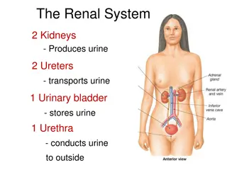
Radiology Imaging Modalities for Renal System Evaluation
Various radiology modalities such as Ultrasound, X-Ray, MRI, CT, and Nuclear Scans are used to assess the renal system. Each modality has its advantages and disadvantages, with Ultrasound and MRI being radiation-free options, and Nuclear Scans providing functional assessment. Understanding the key features and applications of each imaging technique is crucial for accurate diagnosis and treatment planning in renal pathologies.
Download Presentation
Please find below an Image/Link to download the presentation.
The content on the website is provided AS IS for your information and personal use only. It may not be sold, licensed, or shared on other websites without obtaining consent from the author. Download presentation by click this link. If you encounter any issues during the download, it is possible that the publisher has removed the file from their server.

Presentation Transcript
Renal Block Team 437 Radiology Radiology of the renal system Colour Code Important Doctor s notes Extra explanation Radiology Team 437
Objectives: Modality used for assessment of the urinary system X-ray US CT MRI Nuclear Normal anatomy Common pathologies Kidney Ureter Bladder Urethra Radiology Team 437
Modalities used Radiology Team 437
Ultrasound Pros (Advantages) Portable Inexpensive No ionizing radiation Cons (Disadvantages) Time consuming Operator dependent(depends on the skill of the operator). Image Key: White = stones and calcification. Grey = soft tissue. Black = fluid. -Ultrasound: Sound waves that reflect off dense surfaces, giving us a hyper echoic view of the surface. -Objects with less density appear in gray such as sub tissue -Fluids such as water and urine will not reflect the sound wave -The renal pelvis appears white because it is filled with fat Radiology Team 437
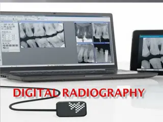
X-RAY Not commonly used on the renal system. Pros (Advantages) Cons (Disadvantages) Inexpensive Ionizing radiation Quick Not definitive Image Key: White = bone and calcification. Grey = soft tissue. Black = air. IVP -Intravenous pyelogram commonly used on the renal system -patient is given contrast which appears bright after entering the renal system IVP -you can tell if there is any stone or obstruction in the ureter -contrast is given Intravenously, and it ends up being excreted by the kidneys Radiology Team 437
MRI CT Stands for (Magnetic Resonance Imaging) Multi leveled X ray, which gives a more definitive and clearer images. Pros (Advantages) Pros (Advantages) No ionizing radiation(uses magnetic fields). Quick A lot of information(can view small structures in the kidney . ( A lot of information(can be used in pregnancy). Cons (Disadvantages) Cons (Disadvantages) Ionizing radiation Time consuming Expensive Image key: same as X ray White = bones and calcification. Grey = soft tissue. Black = air. Expensive Image key: White = high intensity. Grey to black = low intensity. Radiology Team 437
Nuclear scans Pros (Advantages) assess the function Cons (Disadvantages) Time consuming radioactive materials -The patient is given radioactive materials which give off gamma rays, these rays can be detected by special cameras. -This picture shows that the right kidney filtered the radioactive material while the left one did not. Radiology Team 437
Summary -Ultrasound and MRI are the only ones with no ionizing radiation -Nuclear scan is the only one that can asses the function(not only the anatomic structure) Radiology Team 437
Urinary System Anatomy Radiology Team 437
Radiology Team 437
Radiology Team 437
Urinary bladder -Black in Ultrasound(because it s fluid) -Smooth muscle of the bladder -We use it to asses the amount of urine in bladder -Tumors will cause irregularities Radiology Team 437
Common Kidney Pathologies Radiology Team 437
Cysts Cysts: are sac-like structures that may be filled with gas, liquid, or solid materials. It is benign, common and predominantly incidental. Anechoic circular mass , clear borders. Hypo-dense clear border mass in right kidney. -Here it s cyst not tumor, why? Because it has well demarcated fluid inside Radiology Team 437
Stones Radio-opaque (calcium , struvite) (can be seen in X-RAY) Struvite: (magnesium ammonium phosphate) Radio-lucent (uric acid , cysteine) (can t be seen in X-RAY) The best modality for the diagnosis of renal stones is non-contrast CT -Contrast CT will mask the stones because the whole area will become bright -In the other hand non-contrast CT will only make the stones appear bright as you can see in the picture. Radiology Team 437
Stones Uretropelvic junction. Pelvic brim junction: intersection of iliac arteries and ureter -Here we have a stone in the Uretropelvic junction -Here we have a stone in the Pelvic brim junction Radiology Team 437
Hydronephrosis Pyelonephritis It is the infection of the kidney. Acute pyelonephritis results from bacterial invasion of the renal parenchyma. Bacteria usually reach the kidney by ascending from the lower urinary tract. CT scan for a patient with pyelonephritis, we do it only if the patient doesn't respond to the treatment or he had a recurrent pyelonephritis. -A block in the drainage of the renal system which causes the urine to accumulate in the renal pelvis. -When there is a complete obstruction to the ureter by a stone , the kidney eventually fills with urine and become swollen along the ureter -You can notice how the kidney pelvis is dilated or extended if you compare it to the normal ultrasound Radiology Team 437
End-stage renal disease (ESRD) Tumors 1-Benign most common type is angiomyolipoma. -ESRD causes Kidney atrophy -In the picture below we can see atrophy in the left kidney -The right kidney is trying to compensate, that s why it s hypertrophied 2-Malignant most common type is renal cell carcinoma. Radiology Team 437
Congenital kidney diseases Horseshoe Kidney Ectopic Kidney Polycystic Kidney Disease Radiology Team 437
Common Ureter and Urinary bladder Pathologies Radiology Team 437
Ureter pathology vesicoureteral reflux disease -This disease characterized by backflow of the urine -How do we diagnose it ? By giving the patient contrast , after that we will see it go from the ureter back to kidney Radiology Team 437
Urinary bladder pathologies Benign Prostate Hypertrophy Cystitis Image 1: an inflamed urinary bladder (thick surrounding walls). Image 2: This bladder has gas bubbles that could be due to inflammation or infection from gas producing bacteria. Bladder Prostate -Hypertrophied prostate causing the bladder to be compressed Radiology Team 437
Quiz 1)What modality is cheap and with no Ionized radiation? A- Ultrasound B- X-ray C- CT-scan D- MRI 2)What modality is used to assess the function? A- Nuclear scan B- X-ray C- MRI D- CT scan 3)What modality is used with a lot of information and no Ionized radiation? A- X-ray B- Ultrasound C- MRI D- CT scan 4)What type of stones we can see under X-ray? A- Radio-opaque B- Radio-lucent 5)What is the best modality used to diagnose renal stones? A- Contrast CT B- Non-Contrast CT 6)What is the most common type of benign and malignant kidney tumors? A- Transitional cell carcinoma/Renal cell carcinoma B- Angiomyolipoma/Renal cell carcinoma Radiology Team 437 1-A 2-A 3-C 4-A 5-B 6-B
THANK YOU Contact us on: For checking our work. Radiology437@gmail.com @Radiology437 Team Leaders: Rawan Alharbi Faisal Alqusaiyer Team members: Mohammad alasqah Abdulrahaman Altalasi Abdullah Almeaither Abdullah alsergani Abdullah Alomar Mohannad alamri Afnan almustafa Revised by: Homoud al zaid Adel alzahrani Radiology Team 437