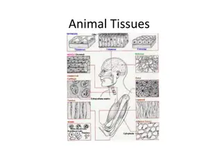
Anatomy of the Larynx: Detailed Illustrations and Muscle Actions
Detailed illustrations and descriptions of the anatomy of the larynx, including structures such as the thyroid cartilage, epiglottis, vocal folds, and muscles like the cricothyroid and posterior cricoarytenoid. The images depict the laryngeal landmarks, ligaments, and nerves, providing a comprehensive insight into the structure and function of the larynx.

Download Presentation

Please find below an Image/Link to download the presentation.
The content on the website is provided AS IS for your information and personal use only. It may not be sold, licensed, or shared on other websites without obtaining consent from the author. Download presentation by click this link. If you encounter any issues during the download, it is possible that the publisher has removed the file from their server.
E N D
Presentation Transcript
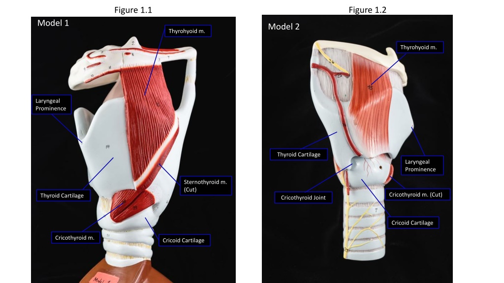
Figure 1.2 Figure 1.1 Model 1 Model 2 Thyrohyoid m. Thyrohyoid m. Laryngeal Prominence Thyroid Cartilage Laryngeal Prominence Sternothyroid m. (Cut) Cricothyroid m. (Cut) Thyroid Cartilage Cricothyroid Joint Cricoid Cartilage Cricothyroid m. Cricoid Cartilage
Figure 1.4 Figure 1.3 Model 2 Model 1 Epiglottis Laryngeal Inlet Epiglottis Aryepiglottic fold Location Vocal Ligament Hyoid Hyoid Corniculate Cartilage Conus Elasticus Piriform Recess Arytenoid Cartilage (mostly covered by muscle) Corniculate Cartilage Cricoid cartilage lamina Arytenoid Cartilage Posterior Cricoarytenoid m. Cricoid cartilage lamina
Figure 1.5 Figure 1.6 Model 2 Epiglottis Laryngeal Inlet Vestibule Ventricle Glottis False (Vestibular) Vocal Fold. (True) Vocal Fold Vocal Ligament Infraglottic Space
Figure 1.7 Internal laryngeal n. Thyrohyoid m. Internal Laryngeal n Piriform Recess Internal Laryngeal n Posterior Cricoarytenoid m. Cricothyroid m. (Cut) Recurrent Laryngeal n. Recurrent Laryngeal n. Recurrent Laryngeal n.
Figure 1.8 Figure 1.9 The blue arrow represents abduction of the vocal fold resulting from posterior cricoarytenoid muscle contraction represented by the yellow arrow. Posterior Cricoarytenoid m. Posterior Cricoarytenoid m.