Female Genital Organs: Ovaries and Anatomy Overview
This informative presentation covers the anatomy of female genital organs, focusing on the ovaries. It explains the functions of the ovaries, their structure, and the various stages of ovarian follicle development. Dr. Sanjay Kumar Bharti's expertise in veterinary anatomy is highlighted through detailed images and descriptions.

Download Presentation

Please find below an Image/Link to download the presentation.
The content on the website is provided AS IS for your information and personal use only. It may not be sold, licensed, or shared on other websites without obtaining consent from the author. Download presentation by click this link. If you encounter any issues during the download, it is possible that the publisher has removed the file from their server.
E N D
Presentation Transcript
Dr. SANJAY KUMAR BHARTI HEAD,VETERINARY ANATOMY
Female Genital Organs Contains following organs 1.-One Pair Gonads 2.- Tubular Organs A.- OVARY. B.- FALLOPIAN TUBE OR OVIDUCT. C.- UTERINE TUBE . D.- VAGINA. E.- URETHRA E.- VULVA, VESTIBULE & CLITORIS F.- Accessory sex glands-.
The ovaries have two functions - "production" and ovulation of oocytes and the production and secretion of hormones. The ovary is attached to the broad ligament by a short fold of peritoneum, called the mesovarium (or ligament of the ovary), through which vessels and nerves pass to the ovary and enter it at the hilus of the ovary.
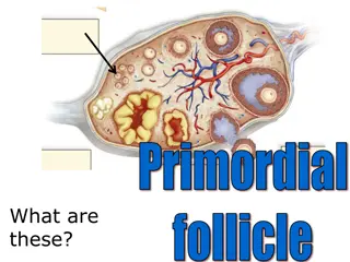
TM-LEM called germinal epithelium. It is continuous with the peritoneal mesothelium. Fibrous connective tissue forms a thin capsule, the tunica albuginea, immediately beneath the epithelium. Like so many other organs the ovary is divided into an outer cortex and an inner medulla. The cortex consists of a very cellular connective tissue stroma in which the ovarian follicles are embedded. The medulla is composed of loose connective tissue, which contains blood vessels and nerves. is lined by cuboidal epithelium, also
In a mature or adult animal, the cortex will contain ovarian follicles, in various stages of development and regressions. Smaller follicles with the ovum surrounded by a layer of follicular cells are located near the periphery. They pass deeper into the ovary as they become larger and develop further but the matured follicle again approaches the surface. The follicle are described as primary, secondary, Graafian and mature Graafian follicles.
Play major roles for transportation of eggs and fertilization takes place. Having 3 parts Infundibulum, Ampula & Isthmus. Mucosa is highly folded i.e.-Primary Secondary and Tertiary types. TM-LEM- Simple Columnar epithelium, Pseudostratifeied Ciliated Columnar in (Ruminanat & Swine) More cila in infundibulum. LMM- absent, LP+SM- Loose CTF with Reticular fibers TMM- I/C,O/L-Thickest in Ampulla. TA/TS-Highly vascular, LCT covered with preritoneal mesothelium isthmus & Thinnest in