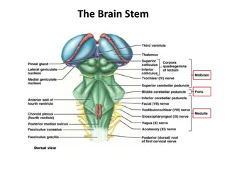
Understanding Internal Structures of the Brain Stem
Explore the intricate details of the brain stem, focusing on the medulla oblongata, pons, midbrain, and the reticular formation. Learn about the distinct components at different levels, such as sensory nuclei, motor decussation, and pathways critical for various functions. Dive into the complexities of the brain stem's internal architecture through detailed examination and visual aids.

Download Presentation

Please find below an Image/Link to download the presentation.
The content on the website is provided AS IS for your information and personal use only. It may not be sold, licensed, or shared on other websites without obtaining consent from the author. Download presentation by click this link. If you encounter any issues during the download, it is possible that the publisher has removed the file from their server.
E N D
Presentation Transcript
Internal Structures of the Brain Stem Please view our Editing File before studying this lecture to check for any changes. Color Code Important Doctors Notes Notes/Extra explanation
Objectives By the end of the lecture, students will be able to : Distinguish the internal structure of the components of the brain stem in different levels and the specific criteria of each level. 1. Medulla oblongata (closed, mid and open medulla) 2. Pons (caudal, mid Trigeminal level and rostral). 3. Mid brain ( superior and inferior colliculi). Describe the Reticular formation (structure, function and pathway) being an important content of the brain stem.
1. Medulla Oblongata Caudal Caudal (closed) (closed) Medulla Medulla o Traversed* by the Central Canal. o Motor Decussation**. o Spinal Nucleus of Trigeminal nerve (Trigeminal sensory nucleus): It is a larger sensory nucleus. It is the brain stem continuation of the Substantia Gelatinosa of spinal cord. * Traversed = travel across or through **Decuss- = crossing Doctor s note: the major thing we see in the closed Medulla is The Motor Decussation. T.S of Caudal part of M.O.
1. Medulla Oblongata Caudal Caudal (closed) (closed) Medulla Medulla Trigeminal Sensory Nucleus & Tract o The Nucleus Extends : Through the whole length of the brain stem and into upper segments of spinal cord. o It lies in all levels of medulla oblongata, medial to the spinal tract of the trigeminal. o It receives pain and temperature from face and forehead (recall this was the function of substantia gelatinosa). o Its tract present in all levels of M.O. is formed of descending fibers that terminate in the trigeminal nucleus.
1. Medulla Oblongata Caudal Caudal (closed) (closed) Medulla Medulla Pyramidal Decussation o Pyramidal decussation is Motor Decussation*. o Formed by pyramidal fibers, (75- 90%) cross to the opposite side o They descend in the lateral white column of the spinal cord as the lateral corticospinal tract. o The uncrossed fibers (the remaining 10-25%) form the ventral corticospinal tract. Extra *Decuss- = crossing
1. Medulla Oblongata Mid Medulla Mid Medulla oTraversed by Central Canal. oLarger size Gracile & Cuneate nuclei, concerned with proprioceptive (knowing the normal body position) deep sensations of the body. oAxons of Gracile & Cuneate nuclei form the internal arcuate fibers; (they form arch like structure) Sensory Decussation.* oPyramids are prominent ventrally. Motor decussation (pyramids): Closed/Caudal Medulla Sensory decussation (internal arcuate fibers): Mid Medulla
Doctors note: dont be confused about Tracts , Fasciculus , Leminiscus . They are all tracts with different locations and shapes. 1. Medulla Oblongata Mid Medulla Mid Medulla After crossing and they ascend and form the medial leminsicus. Sensory Decussation oFormed by the crossed internal arcuate fibers. oMedial Leminiscus*: Composed of the ascending internal arcuate fibers after their crossing. Lies adjacent to the middle line ventral to the central canal Terminates in thalamus. Concerned with proprioceptive deep sensation. At the level of the crossing it is called sensory decussation Before they cross they are the internal arcuate fibers from gracile and cuneate nuclei Medial Lemniscus *lemniscus = ribbon
1. Medulla Oblongata Rostral Rostral (open) Medulla Medulla On the ventral aspect : o The pyramid is clear, with medial lemniscus on either sides of middle line dorsal to the pyramid o Inferior Olivary Nucleus: A convoluted mass of gray matter., lies posterolateral to the pyramids & lateral to the medial leminiscus. It is concerned with the control of movement. The fibers in here will come from the cerebellum. Its dorsal surface forms: Lower part of the floor of the 4th ventricle. o The Inferior Cerebellar Peduncle is, connecting Medulla Oblongata with cerebellum. o Dorsal and lateral to the Inferior cerebellar peduncle lie the Cochlear nuclei (dorsal and ventral). Doctor s Note: we call it open medulla because the central canal will open at this level into the 4thventricle.
1. Medulla Oblongata Rostral Rostral (open) Medulla Medulla Beneath the floor of 4th ventricle lie : 1. Hypoglossal Nucleus. 2. Dorsal Nucleus of Vagus lateral to the hypoglossal nucleus, contains preganglionic parasympathetic fibers. 3. Medial longitudinal fasciculus, it is important association tract, lies close to the midline, ventromedial to the hypoglossal nucleus. Function: Upwards : It links the vestibular nuclei with nuclei of extraocular muscles (in CN 3,4&6) as (vestibulo-ocular tract) to help coordination of eye movements with head movements. Downwards : It links vestibular nuclei with anterior horn cells of spinal cord (cervical & upper thoracic segments) as (vestibulo-spinal tract)---so, the neck & trunk move with head movements.
1. Medulla Oblongata Rostral Rostral (open) Medulla Medulla 4. Vestibular nuclei complex : concerned with equilibrium. Extra 5. Nucleus Ambiguus: (motor nucleus) : lies dorsal to olivary nucleus gives motor fibers along glossopharyngeal N. & vagus N. to motor supply of the constrictors of the pharynx, intrinsic muscles of the larynx & palate. 6. Solitary nucleus (sensory nucleus) : lies ventrolateral to dorsal nucleus of vagus, receive taste sensation from the tongue along the facial (VII), glossopharyngeal (IX) and vagus (X). 7. Tectospinal tract : between tectum of midbrain and spinal cord (involved in head movements during visual and auditory tracking).
2. Pons Caudal Part of Pons Caudal Part of Pons o Divided into an anterior part (Basis Pontis) & a posterior part (Tegmentum) by the Trapezoid Body. o The trapezoid body consists of acoustic fibres (hearing fibers) from cochlear nuclei to ascend into midbrain as lateral lemniscus and terminate in inferior colliculus. o The ventral(anterior) portion is marked by numerous transversely oriented fascicles of pontocerebellar fibres that originate from scattered cell groups, the pontine nuclei(black dots) and that pass to the contralateral side of the cerebellum through the massive middle cerebellar peduncle. Compare: Medial lemniscus Lateral lemniscus Ascending internal arcuate fibers Acoustic fibres from cochlear nuclei Terminates in thalamus Terminate in inferior colliculus
2. Pons Caudal Part of Pons Caudal Part of Pons 5. Deep origin of cranial nerve nuclei : Abducent nucleus Facial motor nucleus 3. The ascending fibres of the medial lemniscus become separated from the pyramid and displaced dorsally. The Medial Lemniscus rotates 90 degrees and lies almost horizontally. 4. Spinal tract & nucleus of Trigeminal. (they become smaller) 2. Bundles of corticospinal & corticonuclear fibres (Pyramidal fibres) 1. Pontine Nuclei: Are small masses of nerve cells, receive corticopontine fibers (involved in motor activity). Their axons form the transverse pontocerebellar fibers which pass to the contralateral side of the cerebellum through Middle Cerebellar peduncles.
2. Pons Level of Trigeminal Nerve Level of Trigeminal Nerve (Mid Pons) o Motor nucleus of the trigeminal nerve: Lies in the lateral part of the floor of the 4thventricle. o Main sensory nucleus of the trigeminal nerve: Reaches its maximum extent in the pons and it lies lateral to the motor nucleus. o Superior cerebellar peduncles form the lateral boundary of the 4thventricle. Rostral Pons Rostral Pons o Superior Medullary Velum: Passes between the two peduncles & forms the roof of the 4thventricle. o Medial longitudinal fasciculus: Lies close to the midline beneath the floor of the 4thventricle. (Mid Pons)
3. Midbrain o It is divided at the level of the cerebral aqueduct into : a dorsal part (Tectum) and a ventral part (Tegmentum) In pons it will be the opposite. o The cerebral aqueduct is surrounded by a pear shaped periaqueductal (central) gray matter. o The most ventral part of the tegmentum is the massive fibrous mass (Crus Cerebri). Extra
3. Midbrain Inferior Colliculus Level Inferior Colliculus Level o Inferior colliculus is a large nucleus of gray matter that lies beneath a corresponding surface elevation. o It is part of the auditory pathway. o It receives fibers from the lateral lemniscus. o Its efferent fibers pass to the thalamus. Extra Structures: 1. Trochlear nucleus: o lies in the central gray matter close to the median plane just posterior to the medial longitudinal bundle. o The fibers of the trochlear nerve decussate in the superior medullary velum. 2. Decussation of the superior cerebellar peduncles in the mid line.
3. Midbrain Inferior Colliculus Level Inferior Colliculus Level Image result for youtube 08:59 Mask Face Extra Flexion of the Trunk 3. Substantia nigra : o Occupies the most ventral part of the tegmentum. o It consists of pigmented, melanin containing neurones. o It projects to the basal ganglia. Its degeneration is associated with Parkinson s disease*. Tone of the muscle will be lost. 4. Ascending Leminisci: o Composed Of: Medial lemniscus. Spinal (Lateral & anterior spinothalamic tracts) Trigeminal (Lateral & medial). Lateral lemniscus. Pill-Rolling Tremors * Slow Shuffling Feet movement
3. Midbrain Crus Crus Cerebri Cerebri o It is a massive mass ventral to the substantia nigra. o It consists entirely of descending cortical efferent fibers (Frontopontine, Corticospinal & corticobulbar and Temporopontine Fibres) to the motor cranial nerve nuclei and to anterior horn cells. o Involved in the coordination of movement. o Present in both levels of colliculi (inferior and superior).
3. Midbrain Superior Colliculus Level Superior Colliculus Level *to remember: The eyes are on top so the superior colliculus visual o A large nucleus of gray matter that lies beneath corresponding elevation. o It forms part of the visual reflexes*. o Its efferent fibers go to the anterior horn cells & to cranial nuclei 3, 4, 6, 7 & 11. o It is responsible for the reflex movements of the eyes, head and neck in response to visual stimuli, as in following a moving object or altering the direction of the gaze.
3. Midbrain Superior Colliculus Level Superior Colliculus Level 1. Oculomotor nucleus: o Situated in the central gray matter close to the median plane. o The fibers of the oculomotor nerve passes anteriorly through the red nucleus to emerge on the medial side of the crus cerebri. 2. Red nucleus : o A rounded mass of gray matter that lies in the central portion of the tegmentum. o Its red coloration is due to its vascularity and the presence of an iron containing pigment in the cytoplasm of its neurons. (Important) o It is involved in motor control. Interpeduncular fossa
Reticular Formation (these are important fibers and mostly unnamed) o It is a complex matrix of nerve fibers & small groups of nerve cells that extends throughout the brain stem. o It has a number of important functions i.e. Respiratory and Cardio- vascular centers are located in the medullary and caudal pontine reticular formation. MEDULLA MIDBRAIN PONS
Reticular Formation Reticular Tract Reticular Tract o Reticulo spinal tracts: Influence a muscle tone & posture o Reticular Activating system: Formed of some of the ascending fibers of the reticular formation. They activate the cerebral cortex through the thalamus. Extra
Reticular Formation Reticular Reticular Neurones Neurones o Raphe Nuclei: Midline reticular nuclei. They are serotonergic* (serotonin producing) Its ascending fibers to the cerebral cortex are involved in the mechanisms of sleep. Its descending fibers to the spinal cord are involved in the modulation of Pain. Extra o Locus Ceruleus: Pigmented neurons that lie in the tegmentum of the caudal midbrain & rostral pons It is the main noradrenergic cell group of the brain. Helps in arousal and sleep-wake cycles. *A serotonergic or serotonergic agent is any chemical that modifies the effects of serotonin in the body
Caudal 1. Trapezoid body (divides it into basis pontis and tegmentum) Transverse pontocerebellar fibers Pontine nuclei Bundles of corticospinal & corticonuclear fibers. Medial lemniscus Spinal tract & nucleus of trigeminal Deep origin of CN 6 & 7 Summary of levels and structures: 2. 3. 4. 5. 6. 7. Caudal / Closed 1. 2. 3. Traversed by central canal Motor decussation Trigmenial sensory nucleus Pons Mid 1. 2. 3. 4. 5. Traversed by central canal Gracile and cuneate nuclei Internal arcuate fibers Sensory decussation Medial leminiscus Mid (level of trigeminal) 1. 2. 3. Motor nucleus of trigeminal Main sensory nucleus of the trigeminal Superior cerebellar peduncle (forms lateral boundary of 4thventricle) Rostral 1. 2. Superior medullary velum Medial longitudinal fasciculus Medulla Rostral / Open 1. Dorsal surface forms lower part of floor of 4thventricle Cochlear nuclei (dorsal and ventral) Hypoglossal nucleus Dorsal nucleus of vagus Medial longitudinal fasciculus Vestibular nuclei complex Nucleus ambiguus Solitary nucleus Tectospinal tract Inferior colliculi (auditory) 1. 2. Trochlear nucleus Decussation of cerebellar peduncle sin the midline Substantia nigra (parkinsons) Ascending lemnisci 2. 3. 4. 5. 6. 7. 8. 9. 3. 4. Midbrain Crus cerebri 1. (present in superior and inferior colliculi) Descending cortical efferent fibers Superior colliculi (visual) 1. 2. Occulomotor nucleus Red nucleus
MCQs MCQs 1. Most axons of cochlear nuclei cross the midline of pons forming: A- the medial lemniscus B- the red nucleus C- trapezoid body D- the medial longitudinal fasciculus Answer: C 5. Solitary nucleus is responsible for which of the following? A- hearing B- taste sensation C- vision D- fine touch Answer: B 2. The axons of the cochlear nuclei are represented in: A- trapezoid body B- medial longitudinal bundle C- tectospinal tract D- spinal lemniscus Answer: A 6. The caudal pons give have deep nuclei of which nerve? A- vagus B- facial C- trigeminal Answer: B 7. The reticular formation is found in: A- midbrain B- pons C- medulla oblongata D- all of the above Answer: D 3. Which of the following lies in the tegmentum of the midbrain: A- oculomotor nuclei B- trochlear nucleus C- red nucleus D- fascial nucleus Answer: C 8. Which nerve fibers emerge on the medial side of the crus cerebri: A- occulomotor B- opthalmic C- vestibulocochlear Answer: A 4. Parkinsons disease results from degeneration of: A- red nucleus B- substantia nigra C- inferior olivary nucleus Answer: B
Members: Alanoud Abuhaimed Leaders: Nawaf AlKhudairy Jawaher Abanumy Image result for google form icon Feedback Feedback anatomyteam436@gmail.com References: 1- Girls & Boys Slides 2- Greys Anatomy for Students 3- TeachMeAnatomy.com @anatomy436 Anatomy Team Image result for youtube Anatomy Team