Understanding the Blood Supply of the Brain


Blood supply of Brain
Essam Ealdin Abdelhady
Salama

Arterial supply of the brain
Four arteries
enter into the
cranial cavity to
supply the brain;
Two vertebral
arteries.
Two internal
carotid arteries.
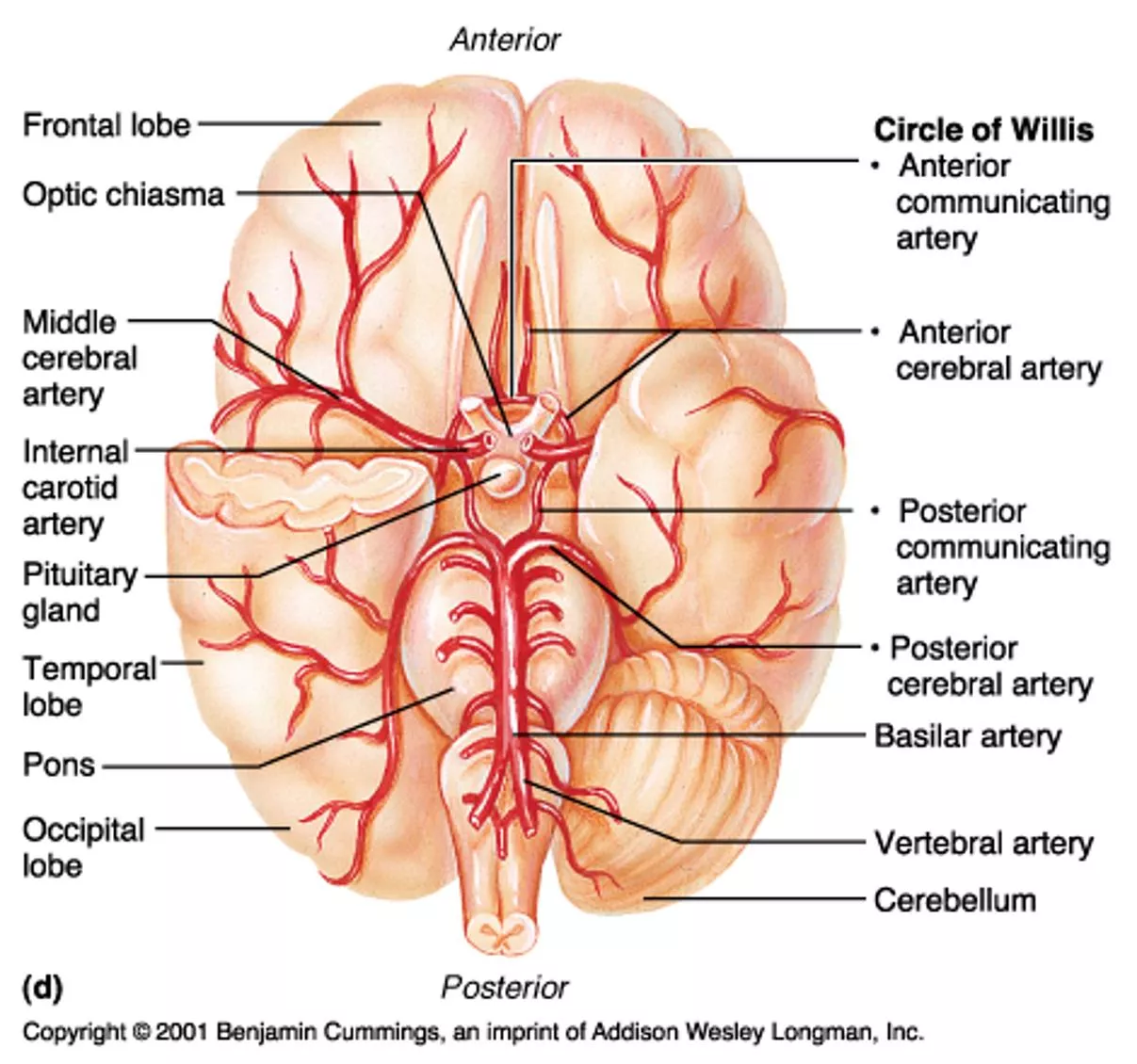


Vertebral arteries.
Aries from 1
st
part of the
subclavian artery.
Entering the cranial cavity
through foramen magnum .
Ventral to pons the
vertebral arteries unite to
form the basilar artery.
Branches;
o
Anterior spinal
o
Posterior spinal.
o
Medullar.
o
Posterior inferior cerebellar.
Basilar artery
Ascends in the basilar
groove of pons.
Ends at upper border of pons
by dividing into two posterior
cerebral arteries.
Branches;
o
Pontine .
o
Labyrinthine .
o
Anterior inferior cerebellar.
o
Superior cerebellar .
o
Posterior cerebral
.
Internal carotid artery
Entering the cranial cavity
through the carotid canal
On the lateral side of optic
chiasma terminating into
o
Anterior cerebral artery
o
Middle cerebral artery.
Before termination giving
o
Posterior communicating .
o
Anterior choroidal.
Circle of Willis
Hexagonal arterial circle lies
on the base of the brain
It is formed by;
Anterior communicating.
Proximal portion of
anterior cerebral.
Internal carotid.
Posterior communicating.
Proximal portion of
posterior cerebral.
Branches of circle of Willis
Central branches
Anteromedial group
Posteromedial group
Poaterolateral group
Anterolateral group
Choroidal
Cortical branches
Anterior cerebral artery
Middle cerebral artery
Posterior cerebral artery
Anterior
cerebral artery
One of two terminal
branches of internal
carotid.
It runs on the medial
surface above corpus
callosum.
Cortical branches
supplying;
Medial part of orbital
surface
Media surface except
occipital lope.
Upper inch of
superiolateral surface.
Middle
cerebral artery
One of two terminal
branches of internal
carotid.
It runs in the stem of the
lateral sulcus.
Cortical branches
supplying;
Lateral part of orbital
surface
Temporal pole
Superiolateral surface
except upper and lower
inches, occipital lobe.
Posterior
cerebral artery
One of two terminal
branches of basilar artery.
It runs on the side of mid
brain.
Cortical branches
supplying;
Tentorial surface.
All aspects of occipital
lope.
Lower inch of
superiolateral surface.
Central group
Consist of :
-Anteromedial group
-Posteromedial group
-Poaterolateral group
-Anterolateral group
-Choroidal a.
Anteromedial branches
: supply
H
ypothalamus
.
P
reoptic area
.
S
upraoptic area
.
Posteromedial branches
Suppl
y;
P
ituitary gland
.
H
ypothalamus
.
T
halamus
.
M
idbrain
.
Posterolateral
(
thalamogeniculate artery
)
supply
P
osterior half of
thalamus
.
G
eniculate body
.
P
ulvinar
.
L
ateral nuclear
group
.
M
ost of ventral
thalamic nuclei
.
Anterolateral branches
(
striate artery
)
Medial striate
arteries;
supply to
;
H
ead of caudate nucleus
.
P
utamen
.
I
nternal capsule
.
Lateral striate a. supply to
C
audate
.
P
utamen
.
G
lobus pallidus
.
I
nternal capsule
.
Choroid arteries
Anterior choroidal artery
: supply to
choroid plexus in lateral
ventricle
.
hippocampal formation
.
globus pallidus
.
posterior limb of internal
capsule
.
amy
g
daliod nucleus
.
tail of caudate nucleus
.
ventrolateral
part
of
thalamus
.
Choroid arteries
Pos
terior choroidal artery
Supply to
T
halamus
.
C
horoid plexus of
third
and lateral
ventricle
.
Blood supply to Corpus striatum
Striatum
: from lateral striate
a.
Head of caudate
: from medial striate a.
Tail of caudate and putamen
: from
anterior choroidal a.
Globus pallidus
: from lateral striate
a
,
anterior choroidal a. And posterior
communicating a.
Blood supply to internal capsule
Anterior cerebral
Middle cerebral
Anterior choroid
Arterial supply of
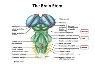
the Midbrain, Pons & Medulla
Midbrain
: Branches from basilar a.
Pons and Medulla
: Anterior spinal a.,
Posterior spinal a., Posterior inferior
cerebellar a., basilar a. and Vertebral a.
Cerebral veins and Venous sinus
Venules in brain form venous plexus in pia
mater
.
Then drain to superficial vein in subarachnoid
space
.
And drain to dural venous sinus
.
Classification of cerebral vein
Superficial cerebral veins
Superior cerebral veins
Inferior cerebral veins
Superficial middle cerebral veins
Deep cerebral veins
Internal cerebral vein
Basal vein (Rosenthal)
Great cerebral vein (Of Galen)
The brain receives its blood supply from four major arteries - two vertebral arteries and two internal carotid arteries. The vertebral arteries branch into the basilar artery, which gives rise to various important branches. The internal carotid arteries supply the anterior and middle cerebral arteries in addition to other crucial branches. The Circle of Willis, a hexagonal arterial circle at the base of the brain, plays a key role in ensuring consistent blood flow. This comprehensive guide details the arterial pathways and branches supplying the brain, shedding light on the intricate network that sustains its vital functions.

Download Presentation

Please find below an Image/Link to download the presentation.
The content on the website is provided AS IS for your information and personal use only. It may not be sold, licensed, or shared on other websites without obtaining consent from the author. Download presentation by click this link. If you encounter any issues during the download, it is possible that the publisher has removed the file from their server.
E N D
Presentation Transcript
Blood supply of Brain EssamEaldinAbdelhady Salama
Arterial supply of the brain Four arteries enter into the cranial cavity to supply the brain; Two vertebral arteries. Two internal carotid arteries.
Vertebral arteries. Aries from 1st part of the subclavian artery. Entering the cranial cavity through foramen magnum . Ventral to pons the vertebral arteries unite to form the basilar artery. Branches; oAnterior spinal oPosterior spinal. oMedullar. oPosterior inferior cerebellar.
Basilar artery Ascends in the basilar groove of pons. Ends at upper border of pons by dividing into two posterior cerebral arteries. Branches; oPontine . oLabyrinthine . oAnterior inferior cerebellar. oSuperior cerebellar . oPosterior cerebral.
Internal carotid artery Entering the cranial cavity through the carotid canal On the lateral side of optic chiasma terminating into oAnterior cerebral artery oMiddle cerebral artery. Before termination giving oPosterior communicating . oAnterior choroidal.
Circle of Willis Hexagonal arterial circle lies on the base of the brain It is formed by; Anterior communicating. Proximal portion of anterior cerebral. Internal carotid. Posterior communicating. Proximal portion of posterior cerebral.
Branches of circle of Willis Cortical branches Anterior cerebral artery Middle cerebral artery Posterior cerebral artery Central branches Anteromedial group Posteromedial group Poaterolateral group Anterolateral group Choroidal
Anterior cerebral artery One of two terminal branches of internal carotid. It runs on the medial surface above corpus callosum. Cortical branches supplying; Medial part of orbital surface Media surface except occipital lope. Upper inch of superiolateral surface.
Middle cerebral artery One of two terminal branches of internal carotid. It runs in the stem of the lateral sulcus. Cortical branches supplying; Lateral part of orbital surface Temporal pole Superiolateral surface except upper and lower inches, occipital lobe.
Posterior cerebral artery One of two terminal branches of basilar artery. It runs on the side of mid brain. Cortical branches supplying; Tentorial surface. All aspects of occipital lope. Lower inch of superiolateral surface.
Central group Consist of : -Anteromedial group -Posteromedial group -Poaterolateral group -Anterolateral group -Choroidal a.
Anteromedial branches : supply Hypothalamus. Preoptic area. Supraoptic area.
Posteromedial branches Supply; Pituitary gland. Hypothalamus. Thalamus. Midbrain.
Posterolateral (thalamogeniculate artery) supply Posterior half of thalamus. Geniculate body. Pulvinar. Lateral nuclear group. Most of ventral thalamic nuclei.
Anterolateral branches (striate artery) Medial striate arteries; supply to; Head of caudate nucleus. Putamen. Internal capsule. Lateral striate a. supply to Caudate. Putamen. Globus pallidus. Internal capsule.
Choroid arteries Anterior choroidal artery : supply to choroid plexus in lateral ventricle. hippocampal formation. globus pallidus. posterior limb of internal capsule. amygdaliod nucleus. tail of caudate nucleus. ventrolateral part of thalamus .
Choroid arteries Posterior choroidal artery Supply to Thalamus. Choroid plexus of third and lateral ventricle.
Blood supply to Corpus striatum Striatum : from lateral striate a. Head of caudate : from medial striate a. Tail of caudate and putamen : from anterior choroidal a. Globus pallidus : from lateral striate a, anterior choroidal a. And posterior communicating a.
Blood supply to internal capsule Anterior cerebral Middle cerebral Anterior choroid
Arterial supply of the Midbrain, Pons & Medulla Midbrain : Branches from basilar a. Pons and Medulla : Anterior spinal a., Posterior spinal a., Posterior inferior cerebellar a., basilar a. and Vertebral a.
Cerebral veins and Venous sinus Venules in brain form venous plexus in pia mater. Then drain to superficial vein in subarachnoid space. And drain to dural venous sinus.
Classification of cerebral vein Superficial cerebral veins Superior cerebral veins Inferior cerebral veins Superficial middle cerebral veins Deep cerebral veins Internal cerebral vein Basal vein (Rosenthal) Great cerebral vein (Of Galen)


")
")