Understanding the Brain: Stem, Midbrain, Pons, Medulla, and Cerebellum Functions
Explore the complex structures and functions of the brain, including the brain stem, midbrain, pons, medulla, and cerebellum. Discover how these regions play crucial roles in controlling reflexes, movements, and vital functions like respiration and posture. Learn about the thick tracts connecting the cerebellum to the brain stem and the protective mechanisms safeguarding the brain within the skull.
Download Presentation

Please find below an Image/Link to download the presentation.
The content on the website is provided AS IS for your information and personal use only. It may not be sold, licensed, or shared on other websites without obtaining consent from the author. Download presentation by click this link. If you encounter any issues during the download, it is possible that the publisher has removed the file from their server.
E N D
Presentation Transcript
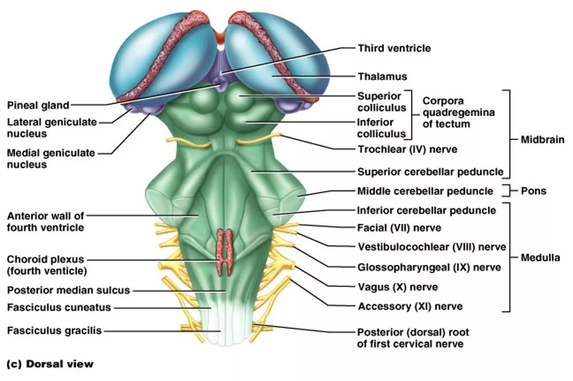
The Brain Stem Midbrain, Pons, Medulla Oblongata
Midbrain Corpora quadrigemina = four bodies (2 pairs) Superior colliculi => nuclei that act in visual reflexes (vision). Inferior colliculi => nuclei that act in auditory reflexes (sound).
The Cerebellum superior view Located dorsal to the pons and medulla Consists of two cerebellar hemispheres Folia
Functions of Cerebellum Located dorsal to the pons and medulla 1) Controls postural reflexes: - coordinates rapid, automatic adjustments of muscles in body to maintain equilibrium. 2) Produces skilled movements:- - implements routines for fine tuned movements. Refines learned routines until the action becomes routine.
* Thick tracts connecting the cerebellum to the brain stem * Superior, Middle and Inferior Cerebellar Peduncles * Fibers to and from the cerebellum are ipsilateral - run to and from the same side of the body
Pons * Contains the pontine respiratory centers. * Contains the nuclei of cranial nerves V, VI, and VII
The Medulla contains: Much of the reticular formation Nuclei influence autonomic functions Vital centers of the reticular formation: 1) Cardiac Control Center 2) Respiratory Control Center 3) Vasomotor Control Center Also contains centers for hiccupping, sneezing, swallowing, vomiting and coughing.
Protection of the Brain The Skull Cranial Meninges Cerebrospinal Fluid Blood-Brain Barrier
Cranial Meninges - 3 layer protective membrane 1.Dura Mater - Composed of two layers: a) Periosteal (Endosteal) outer layer attaches to bone. b) Meningeal inner layer, closer to brain. (These two layers fused, except to enclose the dural sinuses) Composed of dense irregular connective tissue. 2.Arachnoid Layer - spider web-like layer. Composed of loose/fibrous connective tissue. 3. Pia Mater - delicate, follows convolutions. Composed of an outer collagen layer, & inner epithelial elastic and reticular fiber layer.
Cranial Meningeal Spaces Epidural space Potential space superior to dura. Subdural space Potential space between dura and arachnoid mater. Subarachnoid space Filled with CSF Contains the blood vessels supplying brain.
Cerebrospinal Fluid (CSF) Roles of CSF: 1. Cushions and insulates delicate nervous tissue. 2.Gives Buoyancy to the brain ( floats in CSF). 3. Exchange of gases (O2 and CO2), nutrients and wastes. * CSF formed in the choroid plexuses in ventricles of brain
CSF Circulates in: Chambers, Spaces, and the Central Canal.
Blood Brain Barrier (BBB) A restrictive barrier around blood vessels in the brain (created by astrocytes). Prevents most blood-borne toxins from entering the brain but not an absolute barrier. Nutrients such as O2, and glucose can pass. Plus CO2, alcohol, nicotine, and anesthetics. Dopamine? L-Dopa?


