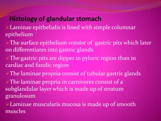
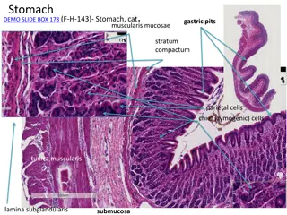
Overview of Salivary Glands Structure
Salivary glands play a crucial role in the production and secretion of saliva, aiding in digestion and oral health. The parotid gland is a compound tubuloacinar gland with serous characteristics, while the sublingual gland is a seromucous type. Minor salivary glands, found in carnivores, vary in composition and distribution. The structure of the glands includes different layers such as stratified squamous epithelium, fibroelastic tissue, and muscular layers.

Uploaded on Oct 03, 2024 | 0 Views
Download Presentation

Please find below an Image/Link to download the presentation.
The content on the website is provided AS IS for your information and personal use only. It may not be sold, licensed, or shared on other websites without obtaining consent from the author. Download presentation by click this link. If you encounter any issues during the download, it is possible that the publisher has removed the file from their server.
E N D
Presentation Transcript
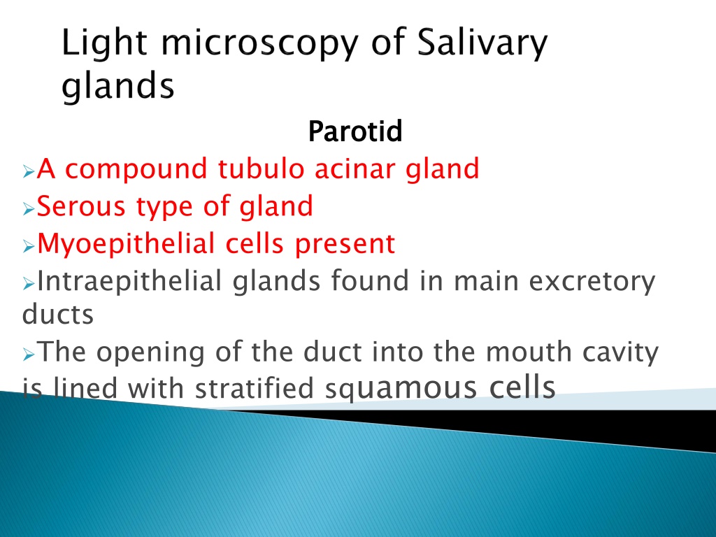
Parotid Parotid A compound tubulo acinar gland Serous type of gland Myoepithelial cells present Intraepithelial glands found in main excretory ducts The opening of the duct into the mouth cavity is lined with stratified squamous cells
Compound tubulo acinar gland Seromucous Myoepithelial cells present Near opening the main duct is lined with Stratified squamous Sublingual salivary gland Seromucous gland Compound tubulo acinar Myoepithelial cells preesent Main duct is lined with stratified cuboidal epithelium Sublingual salivary gland
Found in carnivores Tubuloacinar gland Mucous gland Intercalated and striated ducts are lacking Minor salivary glands Simple May be serous ,mucous or Distributed in surface of of lips and cheek areas Minor salivary glands tubulo or May be serous ,mucous or seromucous Distributed in surface of tongue,vestibular of lips and cheek areas Simple tubulo or tubulo tubulo alveolar glands seromucous glands tongue,vestibular face alveolar glands glands face
Lamina epithelialis is formed by stratified squamous epithelium which may be keratinized or non keratinized depending upon the species Lamina propria submucosae is made up of fibroelastic tissue Laminae muscularis mucosae is absent Tuica muscularis is formed by pharyngeal muscles Tunica adventitia comprised of fibroelastic connective tissue
Laminae epithelialis is made up of stratified squamous ruminants,least in pigs and non keratinized in carnivores Laminae propria is made up of dense fibro elastic connective tissue Laminae muscularis is made up of longitudinally placed smooth muscles Tela submucosa is made up of loose connective tissue and presents seromucous glands epi and is highly keratinized in
In horse the cranial and middle third is made up of striated muscle fiber whereas caudal third is made up of smooth muscle fibers In case of pig the cranial one third is made up of striated striated and smooth muscles and caudal part with smooth muscles Tunica adventitia is made up of loose connective tissue containing blood vessels and lymphatics muscles,middle part is made up of
Non glandular part of non ruminants Tunica mucosae is lined with SSE which is non keratinized in dog Lamina propria made up of loose connective tissue Laminae muscularis mucosae made up of smooth muscles Tela submucosae made up of loose C.T containing submucosal plexus Tunica muscularis made up of smooth muscles Tunica serosa lined with mesothelial cells
Rumen is containing Lamina keratinized granulosum,S.spinosum and stratum corneum Lamina propria is loose and lamina muscularis mucosae is absent Tela submucosa is made up of loose connective tissue and tunica muscularis is made up of smooth muscles epithelialis variety formed by S.corneum,S SSE of
Structures are similar to rumen except the lamina muscularis mucosae which is present at the tip Omasum The omasal laminae presents distinct laminae muscularis mucosae extending from the base to the central core Omasum