Thyroid and Parathyroid Glands Histological Structure Overview

This detailed histological study covers the structure and function of the thyroid and parathyroid glands. It includes information on the stroma, parenchyma, follicular cells, parafollicular cells, and the microscopic structure of the parathyroid gland. The article also delves into the functions of various endocrine cells in the thyroid gland, such as follicular principal cells and parafollicular cells. The parathyroid glands' structure and the secretion of parathyroid hormone are also explained.

Download Presentation

Please find below an Image/Link to download the presentation.
The content on the website is provided AS IS for your information and personal use only. It may not be sold, licensed, or shared on other websites without obtaining consent from the author. Download presentation by click this link. If you encounter any issues during the download, it is possible that the publisher has removed the file from their server.
E N D
Presentation Transcript
Thyroid and Parathyroid glands Red: important. Black: in male|female slides. Gray: notes|extra. Editing file Editing file
OBJECTIVES o Describe the histological structure of THYROID GLAND o Identify and correlate between the different ENDOCRINE CELLS in THYROID GLAND and THEIR FUNCTIONS o The microscopic structure of the PARATHYROID GLAND o The functional structure of the PARATHYROID CELLS Histology team 437 | Endocrine block | Lecture two & three
Thyroid Gland THYROID GLAND STROMA PARENCHYMA 1- Capsule: dense irregular collagenous C.T. 2- Septa (Interlobular septa) 3- Reticular fibers: Thin C.T., composed mostly of reticular fibers with rich capillary plexus surrounds each thyroid follicle. Highly vascular to supply the gland with amino acids and Iodine THYROID FOLLICLES: Are the structural and functional units of the thyroid gland. L/M: 1- Simple cuboidal epithelium: a- Follicular cells. b- Parafollicular cells. 2- Colloid: central colloid-filled lumen. Acidophil gel like material N.B. Each follicle is surrounded by thin basal lamina. Parafollicular cells Follicular cells Colloid Histology team 437 | Endocrine block | Lecture two & three
FOLLICULAR (PRINCIPAL) CELLS 99.9% of the cells PARAFOLLICULAR CELLS (CLEAR CELLS) (C-CELLS) 0.1% of the cells -Pale-stained cells (Clear Cells). -Are found singly or in clusters in between the follicular cells. -Their apices do not reach the lumen of the lumen of the follicle. -Are larger than follicular cells (2-3 times). -Only 0.1% of the epithelial cells. -Have round nucleus -Mitochondria. -RER (moderate). -Well-developed Golgi. L/M -Simple cuboidal cells -Round nucleus with prominent nucleoli. -Basophilic cytoplasm. -Apical surface reaches the lumen of the thyroid follicle. E/M - Mitochondria. - RER synthesis of thyroglobulin - Supranuclear Golgi Complex. - Numerous apically-located lysosomes. - Numerous dispersed small vesicles: contain newly formedthyroglobulin. - Numerous apical short microvilli. Synthesis of thyroid hormones (T4 & T3) Function Secrete calcitonin. Lysosomes are important because it has peroxidase enzyme which convert reduced iodine to Iodine The secretory vesicles are Apical in thyroid gland and basal in parathyroid Calcitonin works with parathyroid hormone to balance the level of Ca++ in the blood Histology team 437 | Endocrine block | Lecture two & three
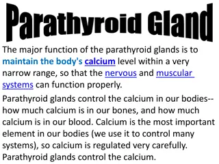
Parathyroid Glands They are 4 glands on the posterior of thyroid gland. Stroma The parenchyma is formed of cords or clusters of epithelial cells (chief cells & oxyphil cell) with blood capillaries in between. These cells are surrounded by reticular fibers. 2- Septa: thin. 3- Reticular C.T. C.T. stroma in older adults often contains many adipose cells. Adipose cells may increase with age - are rich in rER. They secrete parathyroid hormone( blood calcium). Parenchyma 1- Capsule: Each gland has its Thin capsule. Chief cells Oxyphil cells - are slightly eosinophilic. - They are arranged in groups or clusters or as isolated cells. - They are deep eosinophilic (acidophilic) - They have more numerous mitochondria - They are less numerous but larger than chief cells. - They are of unknown function N.B. They may be inactivated chief cells. Histology team 437 | Endocrine block | Lecture two & three
QUESTIONS: QUESTIONS: Q1: What is the epithelium of thyroid follicles? A) Simple columnar epithelium B) Simple squamous epithelium C) Simple cuboidal epithelium D) Simple stratified epithelium Q2: What type of cells is responsible for the secretion of T4 & T3? A) Clear cells B) Principal cells C) Both of them D) None of them 6- B 5- C Q3: What is the structure whose apices do NOT reach the follicle lumen? A) Capsule B) Follicular cells C) Parafollicular cells D) None Q4: What are the cells that can be found singly or in clusters? A) Clear cells B) Principal cells C) None D) All of them 4- A 3- C 2- B 1- C Q5: Which part of thyroid gland stroma, is thin CT with rich capillary plexus? A) Septa B) capsule C) reticular fibers D) none Q6: What is the cell responsible for the secretion of Calcitonin? A) follicular cells B) parafollicular cells C) All of them D) none Histology team 437 | Endocrine block | Lecture two & three
Q7: Which one of the following produce parathyroid hormone? A) Follicular cells B) Parafollicular cell C) chief cells D) oxyphill cells Q8: Which of the following is a Characteristic of oxyphil cells? A) Rich in rER B) abundant mitochondria C) smaller than chief cells D) slightly eosinophilic Q9: Which of the following is a Characteristic of chief cells? A) Rich in rER B) abundant mitochondria C) basophilic D) All of them 11- D 10- B 9- A 8- B Q10: Which of the following is not a histological structure found in parathyroid gland? A) Chief cells B) colloid C) reticular fibers D) oxyphil cells 7- C Q11: Which cell has an un-known function and has numerous mitochondria? A) Follicular cells B) Parafollicular cell C) chief cells D) oxyphill cells Histology team 437 | Endocrine block | Lecture two & three
Team members : Rinad Alghoraiby Ebtesam Almutairi Shahad Alzahrani Fahad Alnuhabi Tareq Allhaidan Abdulmalik Alharbi Team leaders : Khalid Fayez Alshehri Marwah Alkhalil Twitter.com/Histology437 HistologyTeam437@gmail.com


 CELLS")