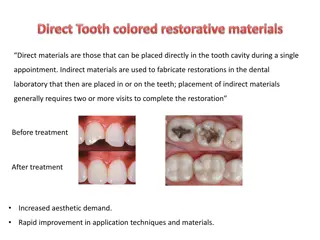
Localization Techniques in Dental Radiography: Enhancing Depth Perception

Dental radiographs, though two-dimensional, can be limited in depicting depth and bucco-lingual relationships. Localization techniques like the right angle and tube-shift methods are used to accurately locate objects such as foreign bodies, impacted or unerupted teeth, and salivary stones within the oral structures. By utilizing these techniques, dentists can enhance their ability to visualize and position objects three-dimensionally.

Download Presentation

Please find below an Image/Link to download the presentation.
The content on the website is provided AS IS for your information and personal use only. It may not be sold, licensed, or shared on other websites without obtaining consent from the author. Download presentation by click this link. If you encounter any issues during the download, it is possible that the publisher has removed the file from their server.
E N D
Presentation Transcript
The dental radiograph is a two dimensional picture of a three dimensional object , a radiograph depict in superio-inferior and anterio- posterior relation ship,so the dental radiograph does not depict the bucco lingual relation ship ,or depth of an object.
There are many times when it is necessary to establish the depth of the structure , such as a foreigh object or impacted tooth within the jaws , LOCALIZATION TECHNIGUES can be be used to obtain this dimensional information , so we can use it to locate the folloing ; 1-Foreign bodies 2-Impacted teeth 3-Unerupted teeth 4-Salivary stone
Types of localization technique 1-Right angle technique 2-Tube-shift technique 3-Stereo radiography 4-Radioopaque media technique The first two technique are the more used because of there simplicity and accuracy
Right angle technique This technique involve the use of at lest two films taken in at right angle to each other , 1-one periapical film is exposed using the proper technique and angulation to show the position of the object in a superrior inferior and anterior posterior relationship
2-An occlusal film is exposed directing the central ray at right angle to the film .the occlusal film shows the object in a bucco- lingual and anterior- posterior relathionship. After the two films have been exposed and processed ,the radiographs are compared to locate the object in three dimensions
Tube shift technique This technique is also called buccal object rule this govering the orientation of structure portrayed in two radiographs exposed at different angulations . One periapical or bite wing film is exposed using proper technique and angulation , A second periapical or bite wing film is then exposed after changing the direction of the x- ray beam . A different horizontal or vertical angulation is used.
For example a different horizontal angulation is used when trying to locate vertically aligned images (root canal), whereas a different vertical angulation is used when trying to locate a horizantallyaligned images (mandibularcanal) after the two films have been exposed and processed , the radiograrhs are compared with each other .
When the dental structure or object seen in the second radiograph appears to have moved in the same direction as the shift of the tubehead,thestructure or object in question is positioned to the lingual For example , if the horizontal angulation is changed by shifting the tube mesially,and the object moves mesiallyon the dental radiograph , then the object lies to the lingual
When the dental structure or object in the second radiograph appears to have moved in the direction opposite the shift of the tube , the object is positioned to the buccal For example, if the horizontal angulation is changed by shifting the tube distally and the object moves mesially on the dental radiograph , the object lies buccal
There is a mnemonic that can be used to remember the buccal object rule SLOB same= Lingual Opposite=Buccal
Stereo radiography this not a widely used because it is time consuming , and the film taken with this technique require a special device , however the operatercan train himselfewithout such device
Radiopaque media Radioopaque media such as barium sulfate lipodol and dionsitecan be used to demarcate cavernous area within hard and soft structure . Such materials are also used to outline soft tissue peripheries such as the profile of the face and neck.