Structure of Gastric Mucosa in Glandular Stomach
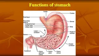
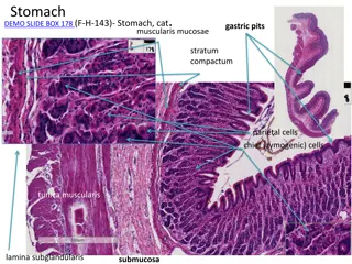
The glandular stomach features laminae epithelialis lined with simple columnar epithelium, leading to differentiation into gastric pits and glands. The laminae propria houses tubular gastric glands, while the laminae muscularis mucosa is composed of smooth muscles. Different regions exhibit distinct gland types, such as cardiac glands in the cardiac area and true gastric glands in the fundic area. Cell types like mucous neck, chief, parietal, and endocrine cells contribute to gland function. The tissue layers include tela submucosa with nerve and vessel components, tunica muscularis with smooth muscle layers, and tunica serosa lined with mesothelium.
- Gastric Mucosa
- Glandular Stomach
- Simple Columnar Epithelium
- Gastrointestinal Anatomy
- Digestive System
Download Presentation

Please find below an Image/Link to download the presentation.
The content on the website is provided AS IS for your information and personal use only. It may not be sold, licensed, or shared on other websites without obtaining consent from the author. Download presentation by click this link. If you encounter any issues during the download, it is possible that the publisher has removed the file from their server.
E N D
Presentation Transcript
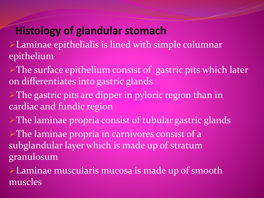
Laminae epithelialis is lined with simple columnar epithelium The surface epithelium consist of gastric pits which later on differentiates into gastric glands The gastric pits are dipper in pyloric region than in cardiac and fundic region The laminae propria consist of tubular gastric glands The laminae propria in carnivores consist of a subglandular layer which is made up of stratum granulosum Laminae muscularis mucosa is made up of smooth muscles
The cardiac area presents cardiac glands distributed in laminae propria which are simple branched coiled tubular gland of mucoussecreting nature The fundic area consist of true gastric glands whose glandular epitheliuum is made up of four varieties of cells mainly mucous neck cell,chief cells,parietal cells and endocrine cells Mucous neck cells,chief cells and parietal cells function likeexocrine glands Mucous neck cells are distributed at the neck and upper part of body of fundic gastric glands and in pig they are also distributed at the base of the gland
The parietal cells are triangular or polyhedral cells which are placed peripheral to the chief cells They are known as Hcl secreting cells and the nucleus is spherical and is basally placed The endocrine cells are known as argentaphin cells or enterochromaffin cells These cells are responsible secretin/gastric inhibitory factors which are transported to the underlying vascularchannel for secretion of
The tela submucosa is made up of loose connective tissue containing submucosal plexus,blood vessels,lymph vessels and lymph nodules Tunica muscularis is made up of smooth muscle cells arranged in innercircularand outer longitudinal layer Tunica serosa is lined with mesothelium
The tunica mucosa is thrown into finger like mucosal folds called intestinal villi In between the two intestinal villi the straight tubular intestinal glands descend in the lamina propria popularly known as glands of liberkuhn The lminae epithelialis is lined with simple columnar epithelium whose epical border consist of numerous microvilli to produce striated border These are responsible to increase the surface area of absorption The surface epithelium presents random distribution of goblet cells which are not distributed at the tipof villi The number of goblet cells increases from cranial to caudal end of small intestine
The glands are lined with undifferentiated columnar cells with few goblet cells.At the base of these glands in case of horse and ruminants there are few specialised pyramidal cells known as Paneth cell The paneth cells are bacteriocidal and are responsible for productionof peptidaseand lysozyme Laminae muscularis mucosae is made up of smooth musclearranged in two layers
These glands open at the base of intestinal villi.In horse the entire duodenum and cranial jejunum consist of Bruner s gland and in ox it is situated in the cranial part of duodenum Ileum presents lymph nodules in form of payer s patches which opens into the lumenof intestine Tunica muscularis is made up of inner circular and outer longitudinal muscles Tunica serosa is lined with mesothelium
Absenceof intestinal villi Absenceof Paneth cells Absenceof submucosal glands Laminae muscularis mucosa notwell developed Large numberof lymph nodules Higher populationof gobletcells Presence of Taenia caeci and Taenia coli in horse and pig which are formed by condensation of outer longitudinal muscle layerof tunica muscularis
