Affections of Salivary Glands in Veterinary Surgery: Overview and Treatment

Salivary gland affections in animals, particularly dogs, are discussed, highlighting the main pairs of salivary glands and their locations. The content covers details on the parotid, mandibular, sublingual, and zygomatic glands, along with their respective ducts. Information on congenital and acquired affections, such as trauma, is provided. Treatment options, including cleaning, debridement, and surgical intervention, are outlined for managing salivary gland conditions in veterinary practice.

Download Presentation

Please find below an Image/Link to download the presentation.
The content on the website is provided AS IS for your information and personal use only. It may not be sold, licensed, or shared on other websites without obtaining consent from the author. Download presentation by click this link. If you encounter any issues during the download, it is possible that the publisher has removed the file from their server.
E N D
Presentation Transcript
AFFECTIONS OF SALIVARY GLAND AND THEIR TREATMENT Regional Veterinary Surgery VSR-421(2+1) Dr. Archana kumari Asstt.Prof. Deptt. of Veterinary Surgery And Radiology
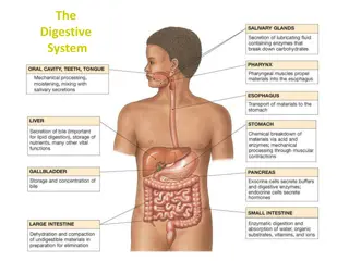
Affections of Salivary Glands and Their Treatment In Dogs:- There are 4 main pairs of salivary glands:- The Parotid, Mandibular, Sublingual, & the Zygomatic glands, which are the dorsal buccal glands in other animals. In horses & ruminants:- There are 3 main pairs of salivary glands:- The Parotid, Mandibular, & Sublingual glands. In Camel:- Parotid, Mandibular and buccal glands. Shape of parotid gland Sheep- Rectangular Buffaloes- Triangular
. Parotid ducts (Stenson s duct) In Bovine parotid ducts open in tobuccal cavity just opposite to upper 5thcheek tooth. In camel it open opposite the fourth cheek tooth below the level of the medial canthus of the eye. The parotid salivary gland is situated ventral to the ear in relation to the caudal border of the mandible. The parotid salivary duct along with the fascial vessels passes ventrally and cranially on the deep face of the caudal part of the mandible and crosses the check superficially just cranial to the massster muscle and ultimately penetrates the cheek near the upper third of fourth cheek tooth. The mandibular (submaxillary) salivary gland is located ventral to the parotid gland just caudal to the mandible. The mandibular savivary duct is located ventral to the parotid gland just caudal to the mandible. The mandibular salivary duct passes forward medial to the mandible to open ventral to the tongue located slightly anterolateral to the frenulum linguae. The sublingual salivary gland is located deep to the mucous membrane along the ventral side of the lateral surface of the tongue near the floor of the mouth. The salivary gland also contains secretary glands like serous, mucous or mixed.
Affection of salivary glands Salivary glands affection can be divided into 2 types:- A. Congenital- Are associated with agenesis or atresia of the parotid ducts, resulting in a fluid-filled swelling proximal to the obstruction site. B. Aquired 1-Trauma Trauma from lacerations or penetrating wounds to the parotid duct or gland is the most common affections associated with the salivary glands. Since the parotid gland and duct is the most superficial, damage to this salivary gland is more commonly seen than to the other salivary glands. If the integrity of the salivary duct or gland is disrupted, there will be increased secretion of saliva from the wound especially during eating.
Treatment Wounds heal spontaneously as with any other wound. Treatment of this condition includes: Cleaning and debridement of necrotic tissue. Fresh wounds can be surgically treated by suturing the parotid duct or suturing the fibrous capsule of the damaged parotid gland or healing by second intention. Provision of liquid diet and avoid noise to reduce salivary secretions. Application of Vaseline to the affected part to prevent excoriation. In horse, nasogastric tube may be used for feeding several days after injury to the parotid gland to prevent stimulation of salivation by eating.
Sialoliths Siololiths or salivary calculi may form in the salivary glands or salivary ducts of the animals. The occurrence of sialoliths or salivary calculi has been reported in different kinds of animals such as dog, cattle, buffaloes, monkeys, donkeys, horses , camels, chimpanzee etc. These are seen more often in horses than in other species. Sialoliths
Siololiths are hard, non-painful, generally smooth and oval in shape, yellowish-grey in colour and moveable swelling that can obstruct usually in rostral portion of the parotid salivary duct in older horses. They are composed of calcium barbonate (80-90%) and organic matter (9-10%). There appears to be no gender or age predisposition to sialolith formation. However, sialolithissis is reported to occur more commonly in animals living in more arid environments
Etiopathogenesis The exact etiopathogenesis of sialolithiasis is not clear, but it is believed that organic matter acts as a nidus around which clacium salts are deposited. The organic matter may be a foreign body, such as grain or grass, entering the parotid duct through the salivary (parotid) papilla, or it may be cellular debris and bacteria as a result of sialodenitis. In the horse, sialoliths may grow to over 10 cm in length and parotid salivary duct obstructionmay occur with large stones, which, in turn, may lead to retention gland dysfunction and oral mucosal ulceration bacteria, resulting in acute sialodenitis, salivary gland dysfunction and oral mucosal ulceration caused by sialolithiasis may occasionally result in discomfort, dysphagia, and possible atrophy of the gland.
Diagnosis The diagnosis of sialolithiasis is based on the following: Clinical signs and palpation. Radiographic examination. Differential diagnosis includes sialodenitis, tooth abscesses, and buccal tumours and metablasia or dystrophic calcification.
Treatment The early surgical intervention is required to save the affected salivary gland from atrophy. Surgical removal of sialoliths by an external approach is traditionally practiced; however, it may cause complications, such as salivary fistula formation if duct closure fails. Consequently, it is recommended to remove them by transoral approach. The incision is made through the buccal mucosa directly over the sialolith. Complications are few with incisions allowed to heal by second intention. Secondary sialodenitis may be a complication of simple sialolith extraction and require postoperative lavage and antimicrobial therapy.
Salivary cyst Salivary cyst is a collection of saliva that has leaked from a damaged salivary gland or salivary duct, and has accumulated in the tissues. In some instances, the duct may have been broken or it may have been bruised with the resultant swelling closing of the duct. Ranula is a mucocele of the sublingual salivary gland and a salivary cyst is aberrant salivary tissue. The large fluid filled swelling will appear similar to a hematoma or seroma. Paracentesis of the swelling yields thick mucoid saliva often mixed with a purulent discharge and rumen card. This condition is almost exclusively seen in dogs, very rarely in cats and occasionally in cattle and buffaloes. Retrograde sialography can confirm diagnosis.
Etiology Trauma such as from choke collars, bite wound or chewing on foreign materials is generally initiating event. Saliva leaking from the torn salivary gland or duct accumulates in the tissue and initiates an intense inflammatory reaction. A connective tissue capsule gradually develops around the saliva to prevent it from migrating further.
Signs In Ranula, there may have some problems in eating and drinking. The animal may limit normal movement of the tongue during drinking and chewing food. In pharyngeal salivary cyst, there may have respiratory difficulty and dysphagia (difficulty in swallowing).
Treatment Sublingual mucocele (Ranula) may be treated with marsupialization (in addition to removal of mandibular and sublingual glands) to facilitate drainage into the mouth. Disscetion and removal of the salivary gland or glands can also be attempted. Continued aspiration of a salivary cyst will not permanently eliminate the problem and sometimes invading bacteria into the cyst can cause an abscess. Chemical ablation of the parotid gland should stop saliva from draining into the cyst. Complete excision of cyst is indicated in high grade infection. Stenson's duct ligation caudal to the placement of the cyst is advocated
Salivary fistula Develops from a wound in the region of a salivary duct to salivary secretion preventing the healing process. A salivary fistula affecting the gland heals in due course of time but in duct involvement, healing will not occur due to continuous flow of saliva through it. Etiology Direct trauma from outside to the duct. Abscess formation in the region leads to rupture of the duct along with communication to outside wound.
Signs:- Dribbling of saliva causing excoriations of the wound. In ruminants, heavy looses of saliva leads to dehydration and indigestion. In few chronic cases, high grade infection of the parotid gland develops. . Treatment:- Salivary fistulas resulting from damaged parotid gland can be treated by reconstructing the duct, transposition or chemical involution of the gland. Surgical repair consists of dissecting over fistula, passing polyethylene catheter caudally towards the glandular part and rostrally through the parotid papilla. A ligature is placed around the duct and brought out this stent through the cheek and keep it in place for several weeks. Chemicals ablation of the gland and duct is accompanied to destroy their functions. Scleresing agents like 10% formalin, 2% iodine, 2% chlrohexidine and 2% silver nitrate may be used for this purpose. The chemical agent especially 10% formalin (because of causing least necrosis and suppurative inflammation) is injected via a canula placed into the parotid duct followed by suturing the canula and duct to prevent leakage during infusion.
Neoplasm Neoplasm of salivary gland are infrequently encountered in animals. In horses, adenocarcinoma, acinar cell tumours, melanomas and mixed cell tumours have all are reported. In canine, a 17 year survey of salivary gland neoplasms in USA revealed adenocarcinoma or carcinomas. Osteosarcome of the mandibular salivary gland is also reported. Clinical signs Parotid swelling and pain that most were Neoplasm
Treatment Treatment of circumscribed and benign neoplasms, excision of the mass is carried out without injuring important nerves and vessels in the vicinity. Surgical excision of mixed cell tumour or acinar cell tumours as well as with melanomas is usually hot successful because of chances of recurrence. Adenocarcinomas often metastasize and it is better not to intervene although, variable success was obtained with surgical excision followed by chemotherapy with cyclophosmamide in dog.
Sub-parotid abscess Sub-parotid abscess generally occurs as a result of strangles in horse and tuberculosis in cattle. A diffuse inflammatory swelling develops which may burst inside or outside if not opened within eight to fourteen days of course. Sometimes, pus materials enter into the larynx and bronchi and results to septic pneumonia or as a fistula of the pharynx may ensue.
Treatment Treatment of this condition is directed to opening the abscess like that of a deep seated abscess without causing injury to important vessels and nerves. Finally, draining of pus material is to be done as per routine procedure. A course of antibiotic for 7 days is indicated.
CANNULATION OF THE STENSONS DUCT Experimental animals for collection of saliva or for parotid sialography. For this purpose, the animal is secured in lateral recumbency and an area just in front of the rostral border of the masseter muscle is prepared for aseptic surgery. A small incision is given along the masseter groove to isolate the parotid duct which lies alongside the facial artery and vein. The duct is then cannulated.
Anaesthesia: Local infiltration with local analgesia Anatomy:Stenson s duct arises from ventro-medial aspect of salivary glands and open at the level of 5th upper tooth. At the site of anterior border of masseter muscle, duct is seen close to masseter muscle and seen as Artery anterior then Vein then Duct (AVD).