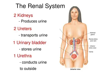
Understanding Urine Concentration Mechanisms in the Renal System
The lecture covers the concepts of urine concentration and dilution in the renal system, focusing on the loop of Henle and countercurrent multiplier and exchange systems. It discusses how the loop of Henle reabsorbs salt and water to determine urine osmolarity, factors influencing medullary gradient formation, and differentiation between water and osmotic diuresis. Clinical implications of conditions like diabetes mellitus and diabetes insipidus are also highlighted.

Download Presentation

Please find below an Image/Link to download the presentation.
The content on the website is provided AS IS for your information and personal use only. It may not be sold, licensed, or shared on other websites without obtaining consent from the author. Download presentation by click this link. If you encounter any issues during the download, it is possible that the publisher has removed the file from their server.
E N D
Presentation Transcript
Urine Concentration and Urine Concentration and Dilution Dilution Red: very important. Green: Doctor s notes. Pink: formulas. Yellow: numbers. Gray: notes and explanation. Physiology Team 436 Renal Block Lecture 8 1 For further understanding please check our Extra Notes file which contains extra explanation from reference books.
Objectives (Please try to read this lecture from the extra notes file and Guyton corners; we have included a few here to help you understand if you already do just skip them) Before starting the lecture, we recommend you to watch this video. Identify and describe that the loop of Henle is referred to as countercurrent multiplier and the loop and vasa recta as countercurrent exchange systems in concentrating and diluting urine. Explain what happens to osmolarity of tubular fluid in the various segments of the loop of Henle when concentrated urine is being produced. Explain the factors that determine the ability of loop of Henle to make a concentrated medullary gradient Differentiate between water diuresis and osmotic diuresis Appreciate clinical correlates of diabetes mellitus and diabetes insipidus 2
Countercurrent system A system in which inflow runs parallel and in close proximity but opposite to the outflow. The operation of such a system allows the outgoing fluid to heat the incoming fluid. The primary function of loop of henle reabsorbs another 20% of the salt/water in tubular fluid that s which determine osmolarity of urine (whether concentrated or diluted) using : Countercutrrent multiplier system. Renal medulla = hyper-osmolarity because of the countercurrent system While collecting duct is where urine concentration is determined, Osmolarity of interstitial fluid in medulla must be high and osmolarity of tubular fluid must be low. 3
Cont. Countercurrent multiplier system: Is the repetitive reabsorption of NaCl by the thick ascending loop of Henle and continued inflow of new NaCl from PCT into loop of Henle. The NaCl reabsorbed from the ascending Loop of henle keeps adding newly arrived NaCl (into LOH from PCT), thus multiplying its concentration in the medulla. Specifically, interstitium of renal medulla continuous flow of Na to meet hyper-osmolarity counter current multiplier system is the most important system of LOH, it detect the conc. of urine by salt/water reabsorption. why is it called multiplier? Because we keep adding more and more of NaCl to medulla, so it's concentration will increase. Guyton corner : [ IMPORTANT ] The most important cause of the high medullary osmolarity is active transport of sodium and co-transport of potassium, chloride, and other ions from the thick ascending loop of Henle into the interstitium. This pump is capable of establishing about a 200-milliosmole concentration gradient between the tubular lumen and the interstitial fluid. Because the thick ascending limb is virtually impermeable to water, the solutes pumped out are not followed by osmotic flow of water into the interstitium. Thus, the active transport of sodium and other ions out of the thick ascending loop adds solutes in excess of water to the renal medullary interstitium. There is some passive reabsorption of sodium chloride from the thin ascending limb of Henle s loop, which is also impermeable to water, adding further to the high solute concentration of the renal medullary interstitium. The descending limb of Henle s loop, in contrast to the ascending limb, is very permeable to water, and the tubular fluid osmolarity quickly becomes equal to the renal medullary osmolarity. Therefore, water diffuses out of the descending limb of Henle s loop into the interstitium and the tubular fluid osmolarity gradually rises as it flows toward the tip of the loop of Henle. 4
Guyton corner : [ IMPORTANT ] Link the figure to the explanation! Steps Involved in Causing Hyperosmotic Renal Medullary Interstitium. Keeping in mind these characteristics of the loop of Henle, let us now discuss how the renal medulla becomes hyperosmotic. First, assume that the loop of Henle is filled with fluid with a concentration of 300 mOsm/L, the same as that leaving the proximal tubule (Figure 28-4, step 1). Next, the active ion pump of the thick ascending limb on the loop of Henle reduces the concentration inside the tubule and raises the interstitial concentration; this pump establishes a 200-mOsm/L concentration gradient between the tubular fluid and the interstitial fluid (step 2). The limit to the gradient is about 200 mOsm/L because paracellular diffusion of ions back into the tubule eventually counterbalances transport of ions out of the lumen when the 200-mOsm/L concentration gradient is achieved. Step 3 is that the tubular fluid in the descending limb of the loop of Henle and the interstitial fluid quickly reach osmotic equilibrium because of osmosis of water out of the descending limb. The interstitial osmolarity is maintained at 400 mOsm/L because of continued transport of ions out of the thick ascending loop of Henle. Thus, by itself, the active transport of sodium chloride out of the thick ascending limb is capable of establishing only a 200-mOsm/L concentration gradient, much less than that achieved by the countercurrent system. Step 4 is additional flow of fluid into the loop of Henle from the proximal tubule, which causes the hyperosmotic fluid previously formed in the descending limb to flow into the ascending limb. Once this fluid is in the ascending limb, additional ions are pumped into the interstitium, with water remaining in the tubular fluid, until a 200-mOsm/L osmotic gradient is established, with the interstitial fluid osmolarity rising to 500 mOsm/L (step 5). Then, once again, the fluid in the descending limb reaches equilibrium with the hyperosmotic medullary interstitial fluid (step 6), and as the hyperosmotic tubular fluid from the descending limb of the loop of Henle flows into the ascending limb, still more solute is continuously pumped out of the tubules and deposited into the medullary interstitium. These steps are repeated over and over, with the net effect of adding more and more solute to the medulla in excess of water; with sufficient time, this process gradually traps solutes in the medulla and multiplies the concentration gradient established by the active pumping of ions out of the thick ascending loop of Henle, eventually raising the interstitial fluid osmolarity to 1200 to 1400 mOsm/L as shown in step 7.Thus, the repetitive reabsorption of sodium chloride by the thick ascending loop of Henle and continued inflow of new sodium chloride from the proximal tubule into the loop of Henle is called the countercurrent multiplier. The sodium chloride reabsorbed from the ascending loop of Henle keeps adding to the newly arrived sodium chloride, thus multiplying its concentration in the medullary interstitium 5
Diluted Urine Has to be: low or no ADH Reabsorb solute not water ADH act on collecting duct to reabsorb water and increase osmolarity 50 mOsm/kg = hypsometric = Diluted Isosmotic fluid from Proximal convoluted tubule Thin descending limb permeable to water , less for NaCl water reabsorbed, tubule osomolality = high in medulla Fluid coming from PCT is isosmotic due to the permeability of Na = water. It won't affect osmolarity. It enter LOH as Isosmotic. In the thin descending limb, which is permeable to water, it start reabsorbing water to be equal to medulla conc. Now the solutes reach the thin ascending limb which is permeable to Nacl . After that they reach the Thick ascending limb which is impermeable to water and permeable to Nacl, the filtrate will be diluted > it will be hyposmotic. Collecting duct will reabsorb Nacl , decreasing the osmolarity more and more ( very hyposmotic = 50 ! ) Thin AL impermeable to water, permeable to NaCl (passive). Volume unchanged , NaCl Thick AL impermeable to water. NaCl reabsorbed (Active) diluting tubule fluid 150 mOsm/kg water. TAL = diluting segment of nephron Collecting duct reabsorb NaCl. osmolality which reach 50 mOsm/kg water 6
Extra - Summary of the Previous Slide Urinary Dilution Mechanism Loop of henle Structure Proximal convoluted tubule Collecting Duct Thin Descending limb Thin Ascending limb Thick Ascending limb Permeable to Both water & NaCl Only to water NaCl Controlled by ADH Impermeable to - Nacl Water ( For the urine to be diluted, there is low or no ADH) Water reabsorption % 65% 20-25% ZERO Reabsorption H2O &NaCl Water (osmosis) NaCl (passive) NaCl (Active) NaCl Osmolarity Isosmotic (300 mOsm/L) Hyperosmotic Hyposmotic Hyposmotic 7
Concentrated Urine Concentration of urine (ADH dependent): (Reabsorbed NaCl in loop of Henle osmolality of interstitium) The first 4 steps are the same of diluted urine TAL impermeable to water. NaCl reabsorbed (Active) diluting tubule fluid 150 mOsm/kg water. Thin AL impermeable to water, permeable to NaCl (passive). Volume unchanged , NaCl Thin descending limb permeable to water .less for NaCl Isosmotic fluid from PCT Fluid reaching CD hypoosmotic (due to urea). ADH causes water to diffuse out up to a max of 300 mOsm/kg water Osmolality of medullary tissue high up to 1200 mOsm/kg water . early CD impermeable to urea ADH allows water reabsorption(passive). NaCl = 600 Urea = 600 Late CD is the site where urea will be absorbed. in step 5, ADH will start to work and absorb water so the conc. of urine will increase The osmolarity in interstituim is high because the change of the osmolarity of the whole body due to ADH How do the kidneys excrete concentrated urine? When ADH levels high urea levels in medullary collecting duct and interstitium equilibrate. Most of water reabsorbed in presence of ADH is in the cortical collecting duct. 8
Sluggish = very slow And the slow blood flow is required for: 1- masticating hyper-osmolarity 2-prevent wash out of solute Vasa Recta Blood supply to loop of henle There are two special features of the renal medullary blood flow that contribute to the preservation of the high solute concentration: The medullary blood flow is low, accounting for less than 5% of the renal blood flow. This sluggish blood flow is sufficient to supply the metabolic needs of the tissues but helps to minimize solute loss from the medullary interstitium. 1. In other words, the vasa recta serve as countercurrent exchanger, minimizing washout of solutes from the medullary interstitium. The vasa recta do not create the medullary hyperosmolarity but they do prevent from being dissipated (very important). 2. Vasa recta maintains hypertonicity by countercurrent exchange. Properties: Recycles NaCl in medulla. Transports H2O from interstitial fluid. Descending limb: Urea transporters - Aquaporin proteins (H20 channels). Ascending limb: Fenestrated capillaries. 1. 2. 3. 4. NaCl and urea diffuse into descending limb and diffuse back into medullary tissue fluid. At each level of the medulla , solute is higher in the ascending limb than in the interstitial fluid and higher in the interstitial fluid than in descending vessels. Walls are permeable to H20, NaCl and urea. Colloid osmotic pressure in vasa recta > interstitial fluid. 10
Extra Difference between counter current systems System Site Function Type of Transport Countercurrent Multiplier Produce medullary hyperosmolaraty In loop of Henle Active transport Countercurrent Exchanger Maintain medullary hyperosmolaraty In vasa recta Passive transport 11
Factors Affecting Urine Concentrations - ADH causes an increase in permeability of DCT, CT, and CD for water. - It also increases the permeability of medullary CD to urea. ADH - The longer the loop of Henle, the greater the countercurrent multiplication effect and more urine concentration. - The new born baby has shorter loop of Henle, which can not concentrate as the adult. Loop of Henle length Delivery of salt to Ascending Loop of Henle Due to a decrease in GFR (Hemorrhage) the filter load of solute is decreased Reabsorption of salt out of ALH decrease in the delivery of salt to ALH resulting in decreased reabsorption of salt from ALH. Delivery of fluid to medullary CT and CD. decreased addition of salts to the medullary ISF ISF : interstitial fluid Medullary blood flow. Urea. decreased medullary longitudinal osmotic gradient and decreased urine concentration. 12
Guyton corner Increased Medullary Blood Flow Guyton corner : Increased Medullary Blood Flow Reduces Urine Concentrating Ability. Certain vasodilators can markedly increase renal medullary blood flow, thereby washing out some of the solutes from the renal medulla and reducing maximum urine concentrating ability. Large increases in arterial pressure can also increase the blood flow of the renal medulla to a greater extent than in other regions of the kidney and tend to wash out the hyperosmotic interstitium, thereby reducing urine concentrating ability. As discussed earlier, maximum concentrating ability of the kidney is determined not only by the level of ADH but also by the osmolarity of the renal medulla interstitial fluid. Even with maximal levels of ADH, urine concentrating ability will be reduced if medullary blood flow increases enough to reduce the hyperosmolarity in the renal medulla. 13
ONLY IN MALES SLIDES Water Diuresis and Osmotic Diuresis Water Diuresis Osmotic Diuresis Increased urine flow rate (no change in solute excretion) Increase in urine flow rate as well as increased solute excretion. Causes: - Excess ingestion of water - Lack of ADH - Defect in ADH receptors in Distal segment of nephron (nephrogenic Diabetes Insipidus) Causes: - Increase plasma glucose level (DM) - Increase level of poorly reabsorbed solutes/ anions - Diuretic drugs (Lasix) Diuresis is mainly due to decrease in water reabsorption in distal segment of nephron. No change to the water reabsorbed proximally Diuresis is mainly due to decrease reabsorption of solute in PCT or LOH. Decrease solute reabsorption results in decrease in water reabsorption proximally as well as distally Increase urine volume results from increased excretion of pure water Increase urine volume results from increased excretion of osmotically active solutes which pulls water with it. Urine osmolality falls far below plasma osmolality. Urine osmolality falls but remains above plasma osmolality. Only about 15% filtered load of water reaching distal segments may remain unabsorbed and excreted in urine (maximum urine volume 20 ml/min) Due to decreased water reabsorption in all segments of nephron, a much greater fraction of filtered water may be excreted (volume more than 20 ml/min) ADH administration will stop diuresis if it is due to lack of ADH or excess ingestion of water. ADH administration will not be effective in Nephrogenic Diabetes Insipidus. ADH administration will NOT stop diuresis. 14
ONLY IN MALES SLIDES Disorders of urinary concentrating ability Cause: inability to produce or release ADH Urine: Low fixed specific gravity(diluted urine). Polyuria (Abnormally large volumes of diluted urine) Polydipsia (Abnormal thirst) Diabetes insipidus Nephrogenic diabetes insipidus Cause: inability of kidney to respond to ADH Diabetes Mellitus Urine: High specific gravity high specific gravity means that urine is concentrated due to glucose excretion in DM 15
Guyton cornerDisorders of Urinary Concentrating Ability Impairment Guyton corner : Disorders of Urinary Concentrating Ability Impairment in the ability of the kidneys to concentrate or dilute the urine appropriately can occur with one or more of the following abnormalities: 1. Inappropriate secretion of ADH. Either too much or too little ADH secretion results in abnormal fluid handling by the kidneys. 2. Impairment of the countercurrent mechanism. A hyperosmotic medullary interstitium is required for maximal urine concentrating ability. No matter how much ADH is present, maximal urine concentration is limited by the degree of hyperosmolarity of the medullary interstitium. 3. Inability of the distal tubule, collecting tubule, and collecting ducts to respond to ADH. Failure to Produce ADH: Central Diabetes Insipidus. An inability to produce or release ADH from the posterior pituitary can be caused by head injuries or infections, or it can be congenital. Because the distal tubular segments cannot reabsorb water in the absence of ADH, this condition, called central diabetes insipidus, results in the formation of a large volume of dilute urine with urine volumes that can exceed 15 L/day. The thirst mechanisms, discussed later in this chapter, are activated when excessive water is lost from the body; therefore, as long as the person drinks enough water, large decreases in body fluid water do not occur. The primary abnormality observed clinically in people with this condition is the large volume of dilute urine. However, if water intake is restricted, as can occur in a hospital setting when fluid intake is restricted or the patient is unconscious (e.g., because of a head injury), severe dehydration can rapidly occur. The treatment for central diabetes insipidusis administration of a synthetic analog of ADH, desmopressin, which acts selectively on V2 receptors to increase water permeability in the late distal and collecting tubules. Desmopressin can be given by injection, as a nasal spray, or orally, and it rapidly restores urine output toward normal. Inability of the Kidneys to Respond to ADH: Nephrogenic Diabetes Insipidus.In some circumstances normal or elevated levels of ADH are present but the renal tubular segments cannot respond appropriately. This condition is referred to as nephrogenic diabetes insipidus because the abnormality resides in the kidneys. This abnormality can be due to either failure of the countercurrent mechanism to form a hyperosmotic renal medullary interstitium or failure of the distal and collecting tubules and collecting ducts to respond to ADH. In either case, large volumes of dilute urine are formed, which tends to cause dehydration unless fluid intake is increased by the same amount as urine volume is increased. Many types of renal diseases can impair the concentrating mechanism, especially those that damage the renal medulla. Also, impairment of the function of the loop of Henle, as occurs with diuretics that inhibit electrolyte reabsorption by this segment, such as furosemide, can compromise urine concentrating ability. And certain drugs, such as lithium (used to treat manic-depressive disorders) and tetracyclines (used as antibiotics), can impair the ability of the distal nephron segments to respond to ADH. Nephrogenic diabetes insipidus can be distinguished from central diabetes insipidus by administration of desmopressin, the synthetic analog of ADH. Lack of a prompt decrease in urine volume and an increase in urine osmolarity within 2 hours after injection of desmopressin is strongly suggestive of nephrogenic diabetes insipidus. The treatment for nephrogenic diabetes insipidusis to correct, if possible, the underlying renal disorder. The hypernatremia can also be attenuated by a low-sodium diet and administration of a diuretic that enhances renal sodium excretion, such as a thiazide diuretic. 16
Guyton corner Summary of Urine Concentrating Mechanism and Changes in Osmolarity in Different Segments of the Tubules Guyton corner : Proximal Tubule. About 65 percent of the filtered electrolytes is reabsorbed in the proximal tubule. However, the proximal tubular membranes are highly permeable to water, so that whenever solutes are reabsorbed, water also diffuses through the tubular membrane by osmosis. Therefore, the osmolarity of the fluid remains about the same as the glomerular filtrate, 300 mOsm/L. Descending Loop of Henle. As fluid flows down the descending loop of Henle, water is absorbed into the medulla. The descending limb is highly permeable to water but much less permeable to sodium chloride and urea. Therefore, the osmolarity of the fluid flowing through the descending loop gradually increases until it is nearly equal to that of the surrounding interstitial fluid, which is about 1200 mOsm/L when the blood concentration of ADH is high. When dilute urine is being formed, owing to low ADH concentrations, the medullary interstitial osmolarity is less than 1200 mOsm/L; consequently, the descending loop tubular fluid osmolarity also becomes less concentrated. This is due partly to the fact that less urea is absorbed into the medullary interstitium from the collecting ducts when ADH levels are low and the kidney is forming a large volume of dilute urine. Thin Ascending Loop of Henle. The thin ascending limb is essentially impermeable to water but reabsorbs some sodium chloride. Because of the high concentration of sodium chloride in the tubular fluid, owing to water removal from the descending loop of Henle, there is some passive diffusion of sodium chloride from the thin ascending limb into the medullary interstitium. Thus, the tubular fluid becomes more dilute as the sodium chloride diffuses out of the tubule and water remains in the tubule.Some of the urea absorbed into the medullary interstitium from the collecting ducts also diffuses into the ascending limb, thereby returning the urea to the tubular system and helping to prevent its washout from the renal medulla. This urea recycling is an additional mechanism that contributes to the hyperosmotic renal medulla. Thick Ascending Loop of Henle. The thick part of the ascending loop of Henle is also virtually impermeable to water, but large amounts of sodium, chloride, potassium, and other ions are actively transported from the tubule into the medullary interstitium. Therefore, fluid in the thick ascending limb of the loop of Henle becomes very dilute, falling to a concentration of about 100 mOsm/L. Early Distal Tubule. The early distal tubule has properties similar to those of the thick ascending loop of Henle, so further dilution of the tubular fluid to about 50 mOsm/L occurs as solutes are reabsorbed while water remains in the tubule 17
Guyton corner Summary of Urine Concentrating Mechanism and Changes in Osmolarity in Different Segments of the Tubules cont. Guyton corner : Late Distal Tubule and Cortical Collecting Tubules. In the late distal tubule and cortical collecting tubules, the osmolarity of the fluid depends on the level of ADH. With high levels of ADH, these tubules are highly permeable to water and significant amounts of water are reabsorbed. Urea, however, is not very permeate in this part of the nephron, resulting in increased urea concentration as water is reabsorbed. This allows most of the urea delivered to the distal tubule and collecting tubule to pass into the inner medullary collecting ducts, from which it is eventually reabsorbed or excreted in the urine. In the absence of ADH, little water is reabsorbed in the late distal tubule and cortical collecting tubule; therefore, osmolarity decreases even further because of continued active reabsorption of ions from these segments. Inner Medullary Collecting Ducts. The concentration of fluid in the inner medullary collecting ducts also depends on (1) ADH and (2) the surrounding medullary interstitium osmolarity established by the countercurrent mechanism. In the presence of large amounts of ADH, these ducts are highly permeable to water, and water diffuses from the tubule into the interstitial fluid until osmotic equilibrium is reached, with the tubular fluid having about the same concentration as the renal medullary interstitium (1200 to 1400 mOsm/L). Thus, a small volume of concentrated urine is produced when ADH levels are high. Because water reabsorption increases urea concentration in the tubular fluid and because the inner medullary collecting ducts have specific urea transporters that greatly facilitate diffusion, much of the highly concentrated urea in the ducts diffuses out of the tubular lumen into the medullary interstitium. This absorption of the urea into the renal medulla contributes to the high osmolarity of the medullary interstitium and the high concentrating ability of the kidney. Several important points to consider may not be obvious from this discussion. First, although sodium chloride is one of the principal solutes that contribute to the hyperosmolarity of the medullary interstitium, the kidney can, when needed, excrete a highly concentrated urine that contains little sodium chloride. The hyperosmolarity of the urine in these circumstances is due to high concentrations of other solutes, especially of waste products such as urea. One condition in which this occurs is dehydration accompanied by low sodium intake. low sodium intake stimulates formation of the hormones angiotensin II and aldosterone, which together cause avid sodium reabsorption from the tubules while leaving the urea and other solutes to maintain the highly concentrated urine. Second, large quantities of dilute urine can be excreted without increasing the excretion of sodium. This is accomplished by decreasing ADH secretion, which reduces water reabsorption in the more distal tubular segments without significantly altering sodium reabsorption.And finally, there is an obligatory urine volume that is dictated by the maximum concentrating ability of the kidney and the amount of solute that must be excreted. Therefore, if large amounts of solute must be excreted, they must be accompanied by the minimal amount of water necessary to excrete them. For example, if 600 milliosmoles of solute must be excreted each day, this requires at least 0.5 liter of urine if maximal urine concentrating ability is 1200 mOsm/L. 18
Summary of Urine Concentrating Mechanism and Changes in Osmolarity in Different Segments of the Tubules cont. Guyton corner : The changes in osmolarity and volume of the tubular fluid as it passes through the different parts of the nephron are shown in Figure 28-8. 19
Thank you! . The Physiology 436 Team: Female Members: Rana Barasain Rania Alessa Ruba Barnawi Male Members: Faisal Alfawaz Abdullah Abuamara Team Leaders: Qaiss Almuhaideb Lulwah Alshiha Contact us: Physiology436@gmail.com @Physiology436 Link to Editing File References: Girls and boys slides. 435 Team. Guyton and Hall Textbook of Medical Physiology (13th Edition). Linda (5th Edition). Special thanks to Team435 s Leaders: Meshal Alhazmy & Khawla Alammari and members! 20