Understanding Renal Physiology: The Role of the Kidneys in Maintaining Homeostasis


Renal Physiology
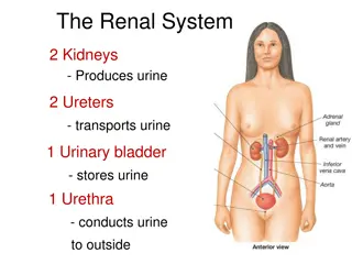
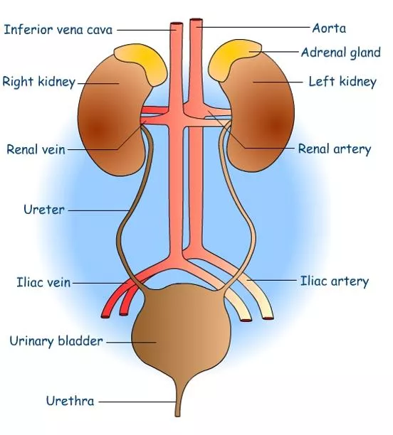
The urinary system is composed of two
kidneys, two ureter, urinary bladder, and
urethra.
The kidney plays a
major role in
controlling the
volume of blood
(water) and also
controlling
ion concentration and PH
. It is the major
excretory organ, responsible for the
removal
of
metabolic waste products from the blood.
The kidneys
act on the plasma first
converting it
to
ultra filtrate
to which it later add and remove
substances so that the
final product is urine.
The kidneys have several functions, including
the following:

1 -
Kidneys
regulate
water and electrolytes balance
: If intake is
less than excretion, the amount of that
substance in the body
will decrease, and vice versa.
2 -
Kidn
e
y
s
r
e
spo
n
sib
l
e
for
e
x
c
r
e
t
i
on
of met
a
bol
i
c
w
a
ste
p
r
odu
c
ts
l
i
ke
u
re
a
f
r
om
the
met
a
bol
i
sm
of
a
m
i
no
ac
ids,
cre
a
t
i
n
i
n
e
f
r
om
muscle
c
r
e
a
t
i
n
e
,
u
r
ic a
c
id
f
r
om
nu
c
leic
ac
id,
b
i
l
iru
b
in
the
e
nd
p
r
odu
c
t
of
Hb b
rea
kdown,
met
a
bol
i
tes
of
v
a
rious
h
o
r
m
o
n
e
s
a
nd
fo
re
i
g
n
c
h
e
m
i
ca
ls
l
ik
e
d
r
u
gs
a
nd
toxins
.
Ho
r
mones
in the
blood
a
re
r
e
moved
in
many
w
a
y
s,
mos
t
ly
in
the
l
i
v
e
r,
but
a
number
of
hormon
e
s
a
re
r
e
moved
in p
a
r
a
l
l
e
l
by
r
e
n
a
l
p
r
o
ce
sses.
The
lev
e
l
of
ni
t
ro
g
e
nous
w
a
ste
in
the
blood
is
t
y
pic
a
l
l
y
e
x
p
re
ssed
a
s
blood
u
re
a ni
t
ro
g
e
n
(
B
U
N
)
.
T
he
u
re
a
c
on
ce
ntr
a
t
i
on
is
norm
a
l
l
y
7
-
18
m
g
/d
L
.
An
a
bnorm
a
l
l
y
e
lev
a
ted
B
UN
is
ca
l
l
e
d
a
z
o
te
m
ia
a
nd
may ind
i
ca
te
r
e
n
a
l
insuf
f
ici
e
n
c
y
.
(See the notes below).
3 -
Kidn
e
y
s
play
e
ssential
role
in
r
eg
ulati
o
n
of
a
rt
e
ri
a
l
p
re
ssure
both
in
long
-
te
r
m
r
eg
ulation
(th
r
ou
g
h
e
x
c
r
e
t
i
on of v
a
ri
a
ble
a
moun
t
s
of sodium
a
nd
w
a
te
r
)
a
nd
in
shor
t
-
te
r
m
r
eg
ulation
(th
r
ou
g
h
s
ec
r
e
t
i
on
of
v
a
soa
c
t
i
ve f
a
c
tors or
subs
t
a
n
ce
s
such
a
s
r
e
nin).
4 -
Kidneys contribute to
acid-base regulation
(along with the
lungs and body buffers) through excreting acids and by
regulating the body buffer stores.
5 -
Kidneys responsible for
regulation
of erythrocyte production
from the bone marrow by
secreting erythropoietin
which
stimulate the bone marrow to produce erythrocytes.
6 -
Kidneys
regulate
1,25-dihydroxy vit. D
3
production
which is
essential in regulation of Ca and phosphate.
7 -
In kidneys,
gluconeogenesis can take place
. Most gluco-
neogenesis occurs in the liver, but a substantial
fraction
occurs in the kidneys, particularly during a prolonged fast.
Anatomy and function of the kidney
Renal function is based on four steps:
1 -
Glomeruli form ultrafiltrate
, which flows
into renal tubules.
2 and 3 -
Tubules reabsorb and secrete
solute and water
from the ultrafiltrate
.
4 -
Tubular fluid
leaves the kidney via the
ureter
to the bladder and out through the
urethra.
B
lood f
r
om
r
e
n
a
l a
r
t
e
ri
e
s
is deliv
e
r
e
d
to
t
he
g
lo
me
ruli. At
one
f
ifth
of
c
a
rd
i
a
c
outpu
t
, th
i
s is
the
hi
g
h
e
st t
i
ssue-spe
c
ific
b
lood fl
o
w
.
The kidney:
The kidney divided into cortex and medulla, and composed of:
[1] Nephron, [
2] Blood vessels
and [
3] Nerves.
[1] The Nephron:
It is a tubular system, and it is
the basic functional unit
of the kidney that
capable of forming
urine by itself
.
There are about 1.3 million nephrons
in each kidney
in human. Kidneys
cannot regenerate new
nephrons
and their number decrease with aging. Each nephron consists of:
A - Bowman’s capsule:
It is the invaginated blind end of the tubule that encased the
glomerulus
(
200µm in diameter and is formed by the invagination of a
tuft of capillaries into the dilated blind end of the nephron
).
The
pressure in the glomerular capillaries is higher than that in other
capillary beds
.
The membrane of the glomerular capillaries is called the
glomerular
membrane
. The
average total area
of each glomerulus is about
0.4
mm
2
, and the
total area of glomerular capillary endothelium
across
which
filtration occurs (i.e. the glomerular membrane) is about
0.8m
2
.
I
n
g
e
n
e
r
a
l,
th
i
s
memb
ra
ne
is
dif
fe
r
e
nt
f
r
om
other
ca
pi
l
la
r
y memb
ra
n
e
s
by
h
a
ving
thr
e
e
la
y
e
rs
ins
t
ea
d
of two
.
Th
e
se
thr
e
e
la
y
e
rs
a
re
e
ndothelial
la
y
e
r
of
the
ca
pi
l
la
r
y
i
t
s
e
lf,
a
b
a
s
e
ment
memb
ra
ne
(b
a
s
a
l
lamina
)
,
a
nd
a
layer of epithelial cells (podocytes)
(As in the figure).
Another type of cells is also present between the basal lamina
and the
endothelium called
mesangial cells
, which are
contractile cells and play a role in the
regulation of glomerular
f
iltration
besides other functions.
Y
e
t,
d
e
spi
t
e
the
numb
e
r
of
l
a
y
e
rs,
the
p
e
rm
e
a
bi
l
i
t
y
o
f
the
g
lo
m
e
rul
a
r
me
m
b
ra
ne
is
f
r
om
10
0
-
500
t
i
mes
a
s
g
re
a
t
a
s
that
o
f
the
u
s
u
a
l
ca
pi
l
la
r
y
.
The
t
r
e
m
e
ndous
p
e
rm
ea
bi
l
i
t
y
of
t
h
e
g
lo
m
e
rul
a
r
memb
ra
n
e
is
ca
used
b
y:
1 -
The
p
re
s
e
n
c
e
of
thous
a
n
d
s
of
small
holes
whi
c
h
a
r
e
ca
l
l
e
d
f
e
n
e
st
r
a
e
in
the
e
ndothelial
ce
l
l
s.
2 -
T
h
e
p
re
s
e
n
c
e
of
large
s
p
a
ce
s
in
t
he
b
a
s
e
ment memb
ra
n
e
.
3 -
I
n
c
ont
i
nui
t
y
of the
ce
l
l
s t
h
a
t
fo
r
m
t
he
e
pi
t
h
e
l
i
a
l
l
a
y
e
r
w
hich
a
r
e
fin
g
e
r-l
i
ke
pro
j
ec
t
i
ons
th
a
t fo
r
ms
s
l
i
ts betw
ee
n
th
e
mselv
e
s c
a
l
l
e
d
sl
i
t
-
p
o
re
s
(filtration slits)
.
B - The Tubule:
Throughout its course, the tubule
is made up of a single layer
of epithelial
cells resting on a basement
membrane. (Note: All epithelial cell layers rest
on a basement membrane).
The structural and immunocytochemical characteristics of these epithelial
cells
vary from segment to segment
of the tubule.
A common feature
is the
presence of tight junctions
between adjacent cells that physically link them
together. The tubule divided in:
1 - Proximal tubules:
It includes
proximal convoluted
tubule and
proximal straight
tubule.
They lie in the
renal cortex
along
with
the
g
lo
m
e
rulus.
The
e
pi
t
h
e
l
i
a
l
ce
l
l
s
of
the
p
r
o
x
i
m
a
l
tubu
l
e
a
re
hi
g
hly
met
a
bol
i
c
ce
l
l
s
,
with
la
r
g
e
number
of
m
i
tochond
r
ia
to
support
e
x
tr
e
mely
ra
pid
ac
t
i
ve
tr
a
nsport
p
r
o
ce
sses
a
nd
they
a
re
in
t
e
rdi
g
i
t
a
ted
with
one
a
nother
a
nd
a
re
uni
t
e
d
by
a
pic
a
l
t
i
g
ht
junction
but
c
ontain
lat
e
r
a
l
in
t
e
r
c
e
l
l
ular
spa
c
e
.
I
t
c
ontains
a
b
r
ush
bord
e
r
due
to the p
re
s
e
n
c
e
of
m
i
c
rovilli.
Reabsorption in the proximal tubule is essentially
isotonic
; i.e. the
osmolality of fluid in all parts of the
proximal tubule is approximately to
that of plasma.
2 - Loops of Henle:
The
nephrons with their glomeruli
located in the
outer portion
of the
renal cortex
have
short loops
of Henle
(cortical nephrons
,
70%),
where
as
those with glomeruli
in the juxtamedullary region of
the cortex
(
juxtamedullary nephrons
,
30%)
have
long loop
extending down into
medullary pyramids.
Loops of Henle
include
: The thin descending segment, the thin
ascending segment, and the thick ascending segment.
T
h
e
t
h
i
n
d
e
s
c
e
n
d
i
n
g
s
e
g
m
e
n
t
o
f
t
h
e
l
o
o
p
o
f
H
e
n
l
e
:
T
h
e
e
p
i
t
h
e
l
i
a
c
e
l
l
s
o
f
i
t
a
r
e
v
e
r
y
t
h
i
n
w
i
t
h
n
o
b
r
u
s
h
b
o
r
d
e
r
a
n
d
v
e
r
y
f
e
w
m
i
t
o
c
h
o
n
d
r
i
a
.
T
h
e
y
a
r
e
h
i
g
h
l
y
p
e
r
m
e
a
b
l
e
t
o
w
a
t
e
r
b
u
t
n
e
a
r
l
y
i
m
p
e
r
m
e
a
b
l
e
t
o
u
r
e
a
,
s
o
d
i
u
m
a
n
d
m
o
s
t
o
t
h
e
r
i
o
n
s
.
A
b
o
u
t
2
0
%
o
f
t
h
e
f
i
l
t
e
r
e
d
w
a
t
e
r
i
s
r
e
a
b
s
o
r
b
e
d
i
n
t
h
e
d
e
s
c
e
n
d
i
n
g
t
h
i
n
l
i
m
b
l
o
o
p
o
f
H
e
n
l
e
The thin ascending segment
of the loop of Henle
: The epithelia cells
of the ascending thin segment, on the other hand,
are far less permeable
to water but more permeable to
urea
and
NaCl
than is the descending
portion
. Because of the ascending thin limb is impermeable to water,
no
water reabsorption
is taking place in this area of the nephron.
The thick ascending segment
of the loop of Henle
:
The epithelial cells of the ascending thick segment
are similar to
those of the proximal tubules
except
that they have a
rudimentary
brush border
and
much tighter
tight junction. The
cells adapted for
strong active transport of
Na,
K,
and
Cl
ions.
On the other hand, the
thick segment is
almost entirely impermeable to both water and
urea.
Therefore,
no water reabsorption is taking place in this area
of
the nephron, and this segment is called
the diluting segment
.
It is the only segment in which active
Cl
pumping normally occur.
This active transport of ions can be inhibited by drugs called loop
diuretics such as frusemide, ethacrynic acid, and bumetanide, which
consequently abolish the intraluminal positivity. Eventually the
passive
absorption of
Na
ions
ceases
.
This thick ascending segment ascends all the way back to the same
glomerulus from which the tubule originated and passes tightly
through the angle between the afferent and efferent arterioles. The
cells of this portion of the thick ascending segment, which are in
complete attachment with the epithelial cells of the afferent and
efferent arterioles, are called
Macula densa.
The specialized smooth muscle cells of the
afferent arterioles
that
come in contact with the macula densa are called
juxtaglomerular
cells
(JG cells) which contain renin granules. Macula densa and JG
cells plus few granulated cells between them are collectively
known as
juxtaglomerular complex or apparatus
which
has a
dense adrenergic neural innervation
.
About 25% of filtered loads of
Na
,
Cl
, and
K
(and other ions such as
Ca
,
HCO
3
-
and
Mg
) are reabsorbed
in the loop of Henle mainly in the
thick ascending limb.
Because the thick segment of the
ascending loop of Henle is
impermeable to water
, most of the
water delivered to this segment
remains in the tubule
, despite the
reabsorption of large amounts of
solute. Thus, the tubular fluid in the
ascending limb
becomes very dilute
as it flows toward the distal tubule
(hypotonic)
.
3 - Distal convoluted tubule:
They lie in the renal cortex.
The distal tubule (also called the
diluting segment
) has almost the same
characteristics as the thick segment of ascending limb of the loop of Henle. It
reabsorbs
Na
ions and other ions but is almost entirely
impermeable to both
water and urea
. This segment is the site of action of special type of diuretics
called thiazide and loop diuretics.
Reabsorption of water
can occurs in the distal tubule but
only
in the presence of
antidiuretic hormone
(ADH, or vasopressin). With high level of ADH, these
tubular segments are permeable to water, but in the absence of ADH, they are
virtually impermeable to water.
Reabsorb Na ions
while
secrete K ions
through increase the activity of
Na-K
ATPase countertransporter
at the basolateral side of the cells under the effect of
the hormone aldosterone.
Reabsorb K
ions while
secrete H ions
via
H-K ATPase countertransporter
at
the luminal border of the cell.
Secretion of H ions
(by
H-ATPase pump
) at the luminal border of the cells after
being generated inside the cell by the action of carbonic anhydrase on water and
CO
2
to form carbonic acid which then dissociates into H ions and HCO
3
-
ions.
Then the available HCO3
-
ions are reabsorbed across the basolateral membrane.
Aldosterone also increases H ion secretion by stimulating the
H-ATPase
pump.
4 - Collecting tubules and ducts:
About eight distal tubules coalesce (unit) to form the collecting
tubule which turns once again away from the cortex and passes
downward into medulla where it becomes the collecting ducts.
The epithelium of collecting ducts is made up of
principal cells
(
P cells
)
which are
involved in Na ions reabsorbtion
and
vasopressin-stimulated water reabsorption and
intercalated cells
(I cells
)
which are
concerned with acid secretion and bicarbonate
transport
.
[B] Blood vessels:
The renal fraction of the total cardiac output is about 21% (vary from
12-30%). In resting adult, the blood flow in renal cortex is about 98%
of the total renal blood flow while in medulla is only 2% of the total
renal blood flow. This is why the O
2
consumption of cortex is much
higher than that of medulla.
Arterial system of the kidney is technically
a portal system
, because
branches twice in the following arrangement: Renal artery
Segmantal artery
Interlobar artery
arcuate artery
Interlobular
artery
Afferent arteriole
branching capillaries in Bowman`s
capsule (glomerulus)
Efferent arterioles
branching around the
tubules so called (Peritubular capillaries)
Venules
Interlobular
veins
arcuate vein
interlobar vein
Renal veins.
Most of the
peritubular capillary network lies in the renal cortex alongside the
proximal, distal, and collecting tubules
.
In the juxtamedullary glomeruli
, long efferent arterioles
extend from the glomeruli down into the outer medulla and
then divide into specialized long and straight capillary loops
called
vasa recta
extended downward into the medulla to lie
side by side with the lower parts of thin segments of
juxtaglomerular loops of Henle
all the way to the renal
papillae. Then, like the loop of Henle, they also loop back
toward the cortex and empty into the cortical veins. This
specialized network of capillaries in the medulla plays an
essential role in the formation of concentrated urine.
[C] Nerve supply:
The kidney has a rich adrenergic sympathetic nerve supply distributed
to the:
1 - Vascular smooth muscle to cause vasoconstriction.
2 - Juxtaglomerular cells to cause renin secretion.
3 - Tubular cells to stimulate Na and water reabsorption.
There is no significant parasympathetic innervation
Glomerular function:
Glomerular filtration rate (GFR):
It is the fluid that filtrate through the glomerulus into Bowman`s
capsule each minute in all nephrons of both kidneys
which is about
125 ml/min
or
180 L/day
in males (10% lower in female). The high
GFR of the glomerular membrane is due to
very high permeability
of
the glomerulus capillaries, which is about
100-500 times as great as
that of the usual capillary.
Yet, despite the tremendous permeability of
the glomerular membrane,
it has an extremely high degree of
selectivity
. The selectivity of the glomerular membrane depends on:
[1] Size of the molecules
:
Neutral substance with effective molecular
diameter of less than 4 nm are freely filtrated, and those with diameter
more than 8 nm (80 A), filtration is zero. Between these two values,
filtration is inversely proportional with diameter.
[2] The electrical charges of the molecules:
This is because the
inner side of the pores of the glomerular membrane is negatively
charged repelling other negatively charged molecules that tend
to pass through pores.
For these two reasons, the glomerular membrane is
almost
completely impermeable
to all plasma proteins
but is highly
permeable
to all other dissolved substances in normal plasma.
The composition of the glomerular filtrate is the same as plasma
except that it has no significant amount of proteins.
The
filtration fraction
is the fraction of the renal plasma flow
that becomes glomerular filtrate. Since the normal plasma flow
through both kidneys is 650 ml/min and the normal GFR is 125
ml/min, the average filtration fraction is about 1/5 or 19%.
Factors that affect GFR
:
GFR is determined by
[1]
the net filtration pressure
and
[2] the
capillary filtration coefficient (K
f
).
GFR can be expressed mathematically as below:
GFR = K
f
X net filtration pressure
Net filtration pressure =
(Glomerular pressure
+
Colloid osmotic pressure in Bowman’s
capsule)
-
(Capsular pressure
+
Colloid osmotic pressure of the
glomerular capillaries)
[1]
The net filtration pressure:
Is
the net pressure forcing fluid through glomerular membrane
(the
Starling forces) which is determined by:
A-
Glomerular capillary hydrostatic pressure:
This can be
affected by several factors:
1 -
Renal blood flow:
Increase blood flow through the nephrons greatly
increases the GFR for two reasons: (A)
The increasing flow increases
the glomerular pressure
which enhances filtration. (B)
The increased
flow through the nephrons allows less time
for plasma proteins to be
more concentrated at the venous end of the glomerular capillaries bed.
Therefore, far less inhibitory influence on glomerular filtration.
2 -
Afferent arteriolar constriction:
Leads
to decrease the rate of blood
flow into the glomeruli
and also
decrease the glomerular pressure and
decrease the GFR
, and vice versa.
3 -
Efferent arteriolar constriction:
A
slight
efferent arteriolar constriction
increases the glomerular pressure causing slight increase in GFR.
However,
moderate and severe
efferent arteriolar constriction causes a
paradoxical decrease in the GFR despite the elevated glomerular
pressure. This is due to the fact that plasma in this case will remain for
long period of time in the glomerulus, and extra large portion of plasma
will filter out. This will increase the plasma colloid osmotic pressure to
excessive level causing a decrease in the GFR.
B -
The change in Bowman’s capsule hydrostatic pressure:
Increasing the hydrostatic pressure in Bowman’s capsule (as in
urinary tract obstruction) reduces GFR and vice versa.
C -
The change in glomerular capillary colloid osmotic pressure:
A decrease in the glomerular capillary colloid osmotic pressure
increases GFR and vice versa.
D -
An increase in the Bowman’s colloid osmotic pressure:
This
may occur in diseases that causes filtration of proteins across
glomerular membrane and consequently increases GFR.
[2] The capillary filtration coefficient (K
f
):
I
s the product of the permeability and filtering surface area of the
capillaries.
It can be affected by:
A -
The changes in the permeability of the glomerular capillaries,
which may be changed in disease state with consequent changes
in the GFR.
B -
The thickness and the surface area of the capillary bed across
which filtration is taking place which can be changed with a
consequent change in the GFR. An example of such change is
contraction or relaxation of mesangial cells in response to various
substances can induce a decrease or an increase in the effective
filtration surface area and eventual changes in the GFR.
In summary
:
GFR
can be affected by:
[1]:
The filtration pressure, which is influenced by:
A. Glomerular capillary hydrostatic pressure which is affected by:
I. Renal blood flow.
II. Afferent arteriolar constriction.
III. Efferent arteriolar constriction.
B. Bowman’s capsule hydrostatic pressure.
C. Glomerular capillary colloid osmotic pressure.
D. Bowman’s colloid osmotic pressure.
[2]:
The capillary filtration coefficient (K
f
), which can be affected by:
A. The permeability of the glomerular capillaries.
B. The thickness and surface area of capillary bed.
The urinary system consists of the kidneys, ureters, urinary bladder, and urethra, playing a crucial role in regulating blood volume, ion concentration, and pH. The kidneys, as the major excretory organ, remove metabolic waste products and help maintain water-electrolyte balance, regulate arterial pressure, acid-base balance, erythrocyte production, and more. Renal function involves glomerular filtration, tubular reabsorption and secretion, ultimately producing urine. Explore the anatomy and functions of the kidneys to understand their essential role in overall health.

Download Presentation

Please find below an Image/Link to download the presentation.
The content on the website is provided AS IS for your information and personal use only. It may not be sold, licensed, or shared on other websites without obtaining consent from the author. Download presentation by click this link. If you encounter any issues during the download, it is possible that the publisher has removed the file from their server.
E N D
Presentation Transcript
Renal Physiology The urinary system is composed of two kidneys, two ureter, urinary bladder, and urethra. The kidney plays a major role in controlling the volume of blood (water) and also controlling ion concentration and PH. It is the major excretory organ, responsible for the removal of metabolic waste products from the blood. The kidneys act on the plasma first converting it to ultra filtrate to which it later add and remove substances so that the final product is urine. The kidneys have several functions, including the following:
1 - Kidneys regulate water and electrolytes balance: If intake is less than excretion, the amount of that substance in the body will decrease, and vice versa. 2 - Kidneys responsible for excretion of metabolic waste products like urea from the metabolism of amino acids, creatinine from muscle creatine, uric acid from nucleic acid, bilirubin the end product of Hb breakdown, metabolites of various hormones and foreign chemicals like drugs and toxins. Hormones in the blood are removed in many ways, mostly in the liver, but a number of hormones are removed in parallel by renal processes. The level of nitrogenous waste in the blood is typically expressed as blood urea nitrogen (BUN). The urea concentration is normally 7-18 mg/dL. An abnormally elevated BUN is called azotemia and may indicate renal insufficiency. (See the notes below).
3 -Kidneys play essential role in regulation of arterial pressure both in long-term regulation (through excretion of variable amounts of sodium and water) and in short-term regulation (through secretion of vasoactive factors or substances such as renin). 4 -Kidneys contribute to acid-base regulation (along with the lungs and body buffers) through excreting acids and by regulating the body buffer stores. 5 -Kidneys responsible for regulation of erythrocyte production from the bone marrow by secreting erythropoietin which stimulate the bone marrow to produce erythrocytes. 6 -Kidneys regulate 1,25-dihydroxy vit. D3production which is essential in regulation of Ca and phosphate. 7 -In kidneys, gluconeogenesis can take place. Most gluco- neogenesis occurs in the liver, but a substantial fraction occurs in the kidneys, particularly during a prolonged fast.
Anatomy and function of the kidney Renal function is based on four steps: 1 - Glomeruli form ultrafiltrate, which flows into renal tubules. 2 and 3 - Tubules reabsorb and secrete solute and water from the ultrafiltrate. 4 - Tubular fluid leaves the kidney via the ureter to the bladder and out through the urethra. Blood from renal arteries is delivered to the glomeruli. At one fifth of cardiac output, this is the highest tissue-specific blood flow.
The kidney: The kidney divided into cortex and medulla, and composed of: [1] Nephron, [2] Blood vessels and [3] Nerves. [1] The Nephron: It is a tubular system, and it is the basic functional unit of the kidney that capable of forming urine by itself. There are about 1.3 million nephrons in each kidney in human. Kidneys cannot regenerate new nephrons and their number decrease with aging. Each nephron consists of:
A - Bowmans capsule: It is the invaginated blind end of the tubule that encased the glomerulus (200 m in diameter and is formed by the invagination of a tuft of capillaries into the dilated blind end of the nephron). The pressure in the glomerular capillaries is higher than that in other capillary beds. The membrane of the glomerular capillaries is called the glomerular membrane. The average total area of each glomerulus is about 0.4 mm2, and the total area of glomerular capillary endothelium across which filtration occurs (i.e. the glomerular membrane) is about 0.8m2. In general, this membrane is different from other capillary membranes by having three layers instead of two. These three layers are endothelial layer of the capillary itself, a basement membrane (basal lamina), and a layer of epithelial cells (podocytes) (As in the figure).
Another type of cells is also present between the basal lamina and the endothelium called mesangial cells, which are contractile cells and play a role in the regulation of glomerular filtration besides other functions. Yet, despite the number of layers, the permeability of the glomerular membrane is from 100-500 times as great as that of the usual capillary. The tremendous permeability of the glomerular membrane is caused by: 1 - The presence of thousands of small holes which are called fenestrae in the endothelial cells. 2 - The presence of large spaces in the basement membrane. 3 - Incontinuity of the cells that form the epithelial layer which are finger-like projections that forms slits between themselves called slit-pores (filtration slits).
B - The Tubule: Throughout its course, the tubule is made up of a single layer of epithelial cells resting on a basement membrane. (Note: All epithelial cell layers rest on a basement membrane). The structural and immunocytochemical characteristics of these epithelial cells vary from segment to segment of the tubule. A common feature is the presence of tight junctions between adjacent cells that physically link them together. The tubule divided in: 1 - Proximal tubules: It includes proximal convoluted tubule and proximal straight tubule. They lie in the renal cortex along with the glomerulus. The epithelial cells of the proximal tubule are highly metabolic cells, with large number of mitochondria to support extremely rapid active transport processes and they are interdigitated with one another and are united by apical tight junction but contain lateral intercellular space. It contains a brush border due to the presence of microvilli. Reabsorption in the proximal tubule is essentially isotonic; i.e. the osmolality of fluid in all parts of the proximal tubule is approximately to that of plasma.
2 - Loops of Henle: The nephrons with their glomeruli located in the outer portion of the renal cortex have short loops of Henle (cortical nephrons, 70%), where as those with glomeruli in the juxtamedullary region of the cortex (juxtamedullary nephrons, 30%) have long loop extending down into medullary pyramids. Loops of Henle include: The thin descending segment, the thin ascending segment, and the thick ascending segment. The thin descending segmentof the loop of Henle: The epithelia cells of it are very thin with no brush border and very few mitochondria. They are highly permeable to water but nearly impermeable to urea, sodium and most other ions. About 20% of the filtered water is reabsorbed in the descending thin limb loop of Henle The thin ascending segmentof the loop of Henle: The epithelia cells of the ascending thin segment, on the other hand, are far less permeable to water but more permeable to urea and NaCl than is the descending portion. Because of the ascending thin limb is impermeable to water, no water reabsorption is taking place in this area of the nephron.
The thick ascending segmentof the loop of Henle: The epithelial cells of the ascending thick segment are similar to those of the proximal tubules except that they have a rudimentary brush border and much tighter tight junction. The cells adapted for strong active transport of Na, K, and Cl ions. On the other hand, the thick segment is almost entirely impermeable to both water and urea. Therefore, no water reabsorption is taking place in this area of the nephron, and this segment is called the diluting segment. It is the only segment in which active Cl pumping normally occur. This active transport of ions can be inhibited by drugs called loop diuretics such as frusemide, ethacrynic acid, and bumetanide, which consequently abolish the intraluminal positivity. Eventually the passive absorption of Na ions ceases.
This thick ascending segment ascends all the way back to the same glomerulus from which the tubule originated and passes tightly through the angle between the afferent and efferent arterioles. The cells of this portion of the thick ascending segment, which are in complete attachment with the epithelial cells of the afferent and efferent arterioles, are called Macula densa. The specialized smooth muscle cells of the afferent arterioles that come in contact with the macula densa are called juxtaglomerular cells (JG cells) which contain renin granules. Macula densa and JG cells plus few granulated cells between them are collectively known as juxtaglomerular complex or apparatus which has a dense adrenergic neural innervation.
About 25% of filtered loads of Na, Cl, and K (and other ions such as Ca, HCO3-and Mg) are reabsorbed in the loop of Henle mainly in the thick ascending limb. Because the thick segment of the ascending loop impermeable to water, most of the water delivered to this segment remains in the tubule, despite the reabsorption of large amounts of solute. Thus, the tubular fluid in the ascending limb becomes very dilute as it flows toward the distal tubule (hypotonic). of Henle is
3 - Distal convoluted tubule: They lie in the renal cortex. The distal tubule (also called the diluting segment) has almost the same characteristics as the thick segment of ascending limb of the loop of Henle. It reabsorbs Na ions and other ions but is almost entirely impermeable to both water and urea. This segment is the site of action of special type of diuretics called thiazide and loop diuretics. Reabsorption of water can occurs in the distal tubule but only in the presence of antidiuretic hormone (ADH, or vasopressin). With high level of ADH, these tubular segments are permeable to water, but in the absence of ADH, they are virtually impermeable to water. Reabsorb Na ions while secrete K ions through increase the activity of Na-K ATPase countertransporter at the basolateral side of the cells under the effect of the hormone aldosterone. Reabsorb K ions while secrete H ions via H-K ATPase countertransporter at the luminal border of the cell. Secretion of H ions (by H-ATPase pump) at the luminal border of the cells after being generated inside the cell by the action of carbonic anhydrase on water and CO2 to form carbonic acid which then dissociates into H ions and HCO3- ions. Then the available HCO3- ions are reabsorbed across the basolateral membrane. Aldosterone also increases H ion secretion by stimulating the H-ATPase pump.
4 - Collecting tubules and ducts: About eight distal tubules coalesce (unit) to form the collecting tubule which turns once again away from the cortex and passes downward into medulla where it becomes the collecting ducts. The epithelium of collecting ducts is made up of principal cells (P cells) which are involved in Na ions reabsorbtion and vasopressin-stimulated water reabsorption and intercalated cells (I cells) which are concerned with acid secretion and bicarbonate transport.
[B] Blood vessels: The renal fraction of the total cardiac output is about 21% (vary from 12-30%). In resting adult, the blood flow in renal cortex is about 98% of the total renal blood flow while in medulla is only 2% of the total renal blood flow. This is why the O2 consumption of cortex is much higher than that of medulla. Arterial system of the kidney is technically a portal system, because branches twice in the following arrangement: Renal artery Segmantal artery Interlobar artery arcuate artery Interlobular artery Afferent arteriole branching capillaries in Bowman`s capsule (glomerulus) Efferent arterioles branching around the tubules so called (Peritubular capillaries) Venules Interlobular veins arcuate vein interlobar vein Renal veins. Most of the peritubular capillary network lies in the renal cortex alongside the proximal, distal, and collecting tubules.
In the juxtamedullary glomeruli, long efferent arterioles extend from the glomeruli down into the outer medulla and then divide into specialized long and straight capillary loops called vasa recta extended downward into the medulla to lie side by side with the lower parts of thin segments of juxtaglomerular loops of Henle all the way to the renal papillae. Then, like the loop of Henle, they also loop back toward the cortex and empty into the cortical veins. This specialized network of capillaries in the medulla plays an essential role in the formation of concentrated urine.
[C] Nerve supply: The kidney has a rich adrenergic sympathetic nerve supply distributed to the: 1 - Vascular smooth muscle to cause vasoconstriction. 2 - Juxtaglomerular cells to cause renin secretion. 3 - Tubular cells to stimulate Na and water reabsorption. There is no significant parasympathetic innervation
Glomerular function: Glomerular filtration rate (GFR): It is the fluid that filtrate through the glomerulus into Bowman`s capsule each minute in all nephrons of both kidneys which is about 125 ml/min or 180 L/day in males (10% lower in female). The high GFR of the glomerular membrane is due to very high permeability of the glomerulus capillaries, which is about 100-500 times as great as that of the usual capillary. Yet, despite the tremendous permeability of the glomerular membrane, it has an extremely high degree of selectivity. The selectivity of the glomerular membrane depends on: [1] Size of the molecules: Neutral substance with effective molecular diameter of less than 4 nm are freely filtrated, and those with diameter more than 8 nm (80 A), filtration is zero. Between these two values, filtration is inversely proportional with diameter.
[2] The electrical charges of the molecules: This is because the inner side of the pores of the glomerular membrane is negatively charged repelling other negatively charged molecules that tend to pass through pores. For these two reasons, the glomerular membrane is almost completely impermeable to all plasma proteins but is highly permeable to all other dissolved substances in normal plasma. The composition of the glomerular filtrate is the same as plasma except that it has no significant amount of proteins.
The filtration fraction is the fraction of the renal plasma flow that becomes glomerular filtrate. Since the normal plasma flow through both kidneys is 650 ml/min and the normal GFR is 125 ml/min, the average filtration fraction is about 1/5 or 19%.
Factors that affect GFR: GFR is determined by [1]the net filtration pressure and [2] the capillary filtration coefficient (Kf). GFR can be expressed mathematically as below: GFR = Kf X net filtration pressure Net filtration pressure = (Glomerular pressure + Colloid osmotic pressure in Bowman s capsule) - (Capsular pressure + Colloid osmotic pressure of the glomerular capillaries) [1]The net filtration pressure: Is the net pressure forcing fluid through glomerular membrane (the Starling forces) which is determined by: A- Glomerular capillary hydrostatic pressure: This can be affected by several factors:
1 - Renal blood flow: Increase blood flow through the nephrons greatly increases the GFR for two reasons: (A) The increasing flow increases the glomerular pressure which enhances filtration. (B) The increased flow through the nephrons allows less time for plasma proteins to be more concentrated at the venous end of the glomerular capillaries bed. Therefore, far less inhibitory influence on glomerular filtration. 2 - Afferent arteriolar constriction: Leads to decrease the rate of blood flow into the glomeruli and also decrease the glomerular pressure and decrease the GFR, and vice versa. 3 - Efferent arteriolar constriction: A slight efferent arteriolar constriction increases the glomerular pressure causing slight increase in GFR. However, moderate and severe efferent arteriolar constriction causes a paradoxical decrease in the GFR despite the elevated glomerular pressure. This is due to the fact that plasma in this case will remain for long period of time in the glomerulus, and extra large portion of plasma will filter out. This will increase the plasma colloid osmotic pressure to excessive level causing a decrease in the GFR.
B - The change in Bowmans capsule hydrostatic pressure: Increasing the hydrostatic pressure in Bowman s capsule (as in urinary tract obstruction) reduces GFR and vice versa. C - The change in glomerular capillary colloid osmotic pressure: A decrease in the glomerular capillary colloid osmotic pressure increases GFR and vice versa. D - An increase in the Bowman s colloid osmotic pressure: This may occur in diseases that causes filtration of proteins across glomerular membrane and consequently increases GFR.
[2] The capillary filtration coefficient (Kf): Is the product of the permeability and filtering surface area of the capillaries. It can be affected by: A - The changes in the permeability of the glomerular capillaries, which may be changed in disease state with consequent changes in the GFR. B - The thickness and the surface area of the capillary bed across which filtration is taking place which can be changed with a consequent change in the GFR. An example of such change is contraction or relaxation of mesangial cells in response to various substances can induce a decrease or an increase in the effective filtration surface area and eventual changes in the GFR.
In summary: GFR can be affected by: [1]: The filtration pressure, which is influenced by: A. Glomerular capillary hydrostatic pressure which is affected by: I. Renal blood flow. II. Afferent arteriolar constriction. III. Efferent arteriolar constriction. B. Bowman s capsule hydrostatic pressure. C. Glomerular capillary colloid osmotic pressure. D. Bowman s colloid osmotic pressure. [2]: The capillary filtration coefficient (Kf), which can be affected by: A. The permeability of the glomerular capillaries. B. The thickness and surface area of capillary bed.


:")