Veterinary Anatomy of Urinary System in Different Animals

The urinary system in animals includes kidneys, ureters, and the bladder, all crucial for filtration and urine excretion. The ureters are excretory ducts from the kidneys, while the bladder stores urine before expulsion. Various diagrammatic structures illustrate the urogenital system of animals, particularly focusing on oxen and cows. The ureters have abdominal and pelvic parts, with specific routes through the body depending on the species and gender.

Download Presentation

Please find below an Image/Link to download the presentation.
The content on the website is provided AS IS for your information and personal use only. It may not be sold, licensed, or shared on other websites without obtaining consent from the author. Download presentation by click this link. If you encounter any issues during the download, it is possible that the publisher has removed the file from their server.
E N D
Presentation Transcript
VETERINARY ANATOMY VETERINARY ANATOMY- -UNIT TOPIC TOPIC- - URINARAY SYSTEM URINARAY SYSTEM- -URETER & URINARY BLADDER URINARY BLADDER- - OF DIFFERENT ANIMALS ANIMALS UNIT- -6 6 URETER & OF DIFFERENT Instructor- DR DR. . SANJAY SANJAY KUMAR KUMAR BHARTI BHARTI HOD, VETERINARY ANATOMY Bihar Veterinary College Patna
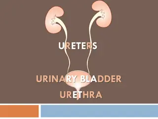
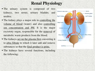
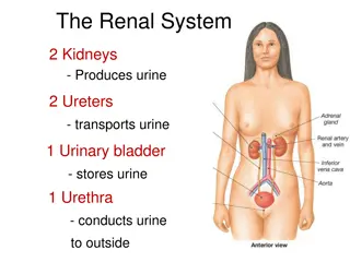
URINARAY SYSTEM URINARAY SYSTEM The urinary organs are the kidneys, ureters, bladder and urethra The kidneys are organs having two supra renal glands play major role for filtration and secrete the urine. The ureters are tubes, which convey the urine to the urinary bladder The urinary bladder is a reservoir for the urine & located on UB The urine accumulates in the bladder and is then expelled through the urethra
DIGRAMATIC Structures of UROGENITAL SYSTEM-OX-COW
DIGRAMATIC Structures of UROGENITAL SYSTEM-OX
DIGRAMATIC Structures of kidney of URINARY SYSTEM OX
URETER -OX The ureters are the excretory ducts of the kidneys and each begins at the junction of the calyces majores and terminates at the bladder It is about 6 to 8 mm in diameter. The right ureter emerges out of the hilus of the right kidney from its ventral face, runs inwards, gains the middle of the medial border of the kidney and runs along it The left ureter emerges out of the hilus on the antero-lateral aspect of the dorsal face, crosses over this face medially and gains the medial border and runs backwards. Each ureter consists of abdominal and pelvic parts
URETER-OX-Contin.. The abdominal part runs backwards and inwards, the right being related to the lateral face of the caudal vena cava and the left to the aorta Then it passes back in the sub-peritoneal tissue on the ventral face of the psoas muscle crosses the external iliac artery and enters the pelvic cavity The pelvic part passes backwards and downwards on the lateral wall of pelvic cavity, turns inward and pierces the superior wall of the bladder near the neck In the male, the pelvic part enters the genital fold and crosses vas deferens whereas in the female it is situated in the dorsal part of the broad ligament of the uterus
Species difference Species difference- -URETER URETER Sheep and goat- It resembles that of ox Horse-The ureter arises from the renal pelvis and real pelvis more dilated - Tube is narrow but longer than the ox. Dog- It resembles that of the horse except origin point. Pig- The Cranial part is wide and gradually diminishes in calibre Slightly flexuous Fowl The ureter is white in colour. It may be divided into a renal part and a pelvic part. The renal part embedded in the ventral surface of the kidney and the pelvic part runs backward in relation to the vas deferens or oviduct It opens into the urodeum of the cloaca, internal to the opening of the vas deferens or oviduct
URETER URETER- - FOWL FOWL
URINARY BLADDER-OX The urinary bladder is a musculo-membranous sac, differs in form, size and position according to the amount of its contents When contracted, it is a dense pyriform mass about the size of a fist and lies on the ventral wall of the pelvic cavity at a variable distance behind the pelvic inlet When moderately full, it is oval in shape and extends into the abdominal floor. Its capacity is about 3 to 4 litres. The bladder has 3 parts-a vertex, a body and neck The cranial part or the vertex is a blind end and presents in its middle a rounded cul-de-sac called as cicatrix - the vestige of the urachus, which forms a connection between the bladder and the allantois in the fetus The middle part or body is rounded and somewhat flattened from above downwards It presents two surfaces- superior and inferior which are convex when full The caudal narrow extremity is the neck and it joins the urethra
URETER AND URINAY BLADDER OF OX-COW
Relations- URINARY BLADDER They vary according to the amount of the contents and sex The ventral surface is related to the floor of the pelvis and extends into the abdomen as it distends The dorsal surface in the male is related to the rectum, the genital fold, and the terminal parts of the vas deferens, the seminal vesicle and the prostate In the female, it is related to the body of uterus and vagina. When the bladder is full, the vertex reaches the rumen and small intestine
Ligaments- URINARY BLADDER The bladder is fixed in position by three peritoneal folds -the ligaments of the bladder The ventral or middle ligament is a median triangular fold extends from the ventral face of the bladder to the floor of the pelvis and abdomen In the newborn animal, it is extensive and reaches to the umbilicus The lateral ligaments are two and extend on either side of the lateral aspects of the bladder to the lateral wall of the pelvis. Each contains in its free edge a round cord the round ligament, which is the remnant of the fetal umbilical artery The caudal part of the bladder has no peritoneal covering and is attached to the surrounding parts by fat and connective tissue. The mucous membrane is pale, thin and is loosely attached to the submucous tissue
Ligaments- URINARY BLADDER It forms numerous folds when bladder is empty and contracted It is modified over a triangular area on its dorsal wall close to neck called the trigonum vesicae where the mucous membrane is firmly attached without any folds The two cranial angles of the trigonum vesicae present the openings of the two ureters and the caudal angle shows the opening of the bladder into the urethra -internal urethral orifice The two urethral orifices are placed near each other on either side of the median line. From each urethral orifice, a fold of mucous membrane passes backward and inward, uniting with its fellow to form a median urethral crest in the first part of urethra. The terminal part of ureter after piercing the muscular coat of the bladder and passes for a distance of about 2.5 cm between the muscular and mucous coats before piercing the latter This arrangement constitutes a valve, which prevents the return of urine from the bladder into ureter The internal urethral orifice lies at the apex of the trigonum
Species difference Species difference- -UB UB Sheep and Goat- Resembles the ox Horse Shorter but wider It does not extend into the abdomen as forward as in the ox Caudal part of the bladder is retro peritoneal Pig It is relatively very large When full, it is abdominal in position Dog When empty, it is pelvic in position and when full it is entirely abdominal in position Fowl The ureters open into the urodeum of the cloaca. So there is no bladder as such
DIGRAMATIC Structures of kidney-Ureter of FOWL.