Understanding Urinary System Function in Veterinary Medicine
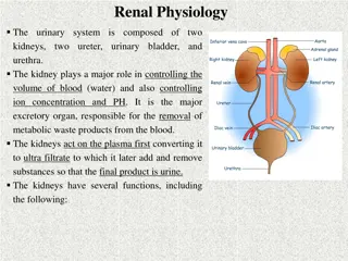
The urinary system in veterinary medicine involves the anatomy and physiological functions of maintaining body fluid composition and volume. Key processes like urine production, filtration, reabsorption, and regulation through nephrons play vital roles in maintaining overall health. This article covers the normal physiological functions of the nephron, urine production factors, and the distinct functions of the glomerular capillary wall, proximal tubule, distal tubule, and loop of Henle in the renal system.

Download Presentation

Please find below an Image/Link to download the presentation.
The content on the website is provided AS IS for your information and personal use only. It may not be sold, licensed, or shared on other websites without obtaining consent from the author. Download presentation by click this link. If you encounter any issues during the download, it is possible that the publisher has removed the file from their server.
E N D
Presentation Transcript
DISEASES OF URINARY SYSTEM Unit-II Veterinary Medicine [ Part-I] By Dr. Anil Kumar Assistant Professor Department of VCC, BVC, BASU, Patna
Anatomy and Physiological Functions Maintaining composition of body fluids the volume and Clean end products Produce erythropoietin, renin angiotensin, and calcitriol the body fluids of hormones such as Have metabolic functions
Urine Production Urine production relies on two factors: Proper function of the nephron Blood supply to the glomerulus Glomerular filtration rate (GFR) of 3 5 mL/min/kg for dogs and 2.5 3.5 mL/min/kg for cats The mean renal arterial pressure is 100 mmHg, for both dogs and cats While the pressure within the glomerular capillary is 55 mmHg in dogs and 59 mmHg in cats. The pressure within the glomerular capillary is substantially higher than the pressure within Bowman s space, thus leading to a difference in hydrostatic pressure of 35 mmHg, facilitating rapid filtration through the glomerular capillary wall
The glomerular capillary wall has both size and charge selectivity, i.e. all molecules<4 nm and positively charged molecules to pass easily through the membrane Filtration is the initial step in urine production via the passive movement of protein and cell free plasma from the glomerular capillaries into Bowman s space to form a plasma ultrafiltrate. The proximal tubule s (Reabsorption): Reabsorption of 50 55% of filtered water and sodium, and virtually all the glucose, phosphate, and amino acids found in the ultrafiltrate Reabsorption of solutes uses mechanisms of active transport, while water moves passively along the concentration gradient as solutes move out of the tubule lumen and into the tubular
The distal tubule : it is relatively impermeable to water but continues to reabsorb sodium and chloride. Parathyroid hormone acts on the distal tubules to regulate calcium reabsorption The loop of Henle: Continues to reabsorb sodium and chloride as well as calcium and magnesium through an active transport mechanism A concentration gradient is no longer established following the removal of solutes (glucose, phosphate, and amino acids) in the proximal tubules. Very little water is reabsorbed in the loop of Henle and causes a dilution of the filtrate and a hyposmotic fluid
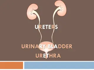
Collecting duct: The fluid reaching the collecting duct is hyposmotic to plasma. Under the influence of antidiuretic hormone (ADH), released from the posterior pituitary, the collecting ducts begin to reabsorb water Fluid in the collecting ducts is deposited in the renal pelvis as urine, where it enters the ureter and finally the bladder, where it awaits micturition The kidneys are highly sensitive organs and can easily be damaged. Kidney receive 20% of the cardiac output, and 90% of that blood goes to the kidney cortex, where key structures of the nephron are exposed to toxins concentrated in the blood.
While autoregulation is used to keep renal blood flow(RBF), and therefore GFR, at a constant level The periods of hypotension (MAP <60 mmHg) can cause an interruption of oxygen delivery to the kidney. Cells of the proximal tubule and thick ascending loop of Henle are particularly sensitive to ischemia as they have a very high metabolic rate
Causes and effects of therenin angiotensin ald osterone system (RAAS)
If the kidneys are damaged, they must lose 66% of their function before clinical signs of renal insufficiency will appear, and 75% before developing renal failure and bloodwork changes Azotemia An abnormal concentration of urea nitrogen and creatinine in the blood Causes: Diminished capacity of the kidney to remove these substances Changes in the volume of blood reaching the kidneys Impaired outflow of urine, AND consequently lead to---Renal failure When azotemia is identified, it is important to distinguish among prerenal, renal, and postrenal causes
Prerenal azotemia: Table:1 DISORDERS ASSOCIATED WITH AZOTEMIA AND INADEQUATE URINE DESPITE NORMAL RENAL FUNCTION IN DOGS AND CATS Any disorder that decreases renal CONCENTRATION perfusion (e.g., dehydration, heart failure, or hypovolemia) or that results in increased production of urea (e.g., gastrointestinal hemorrhage). With few exceptions dogs and cats with purely prerenal causes of azotemia produce concentrated urine (i.e., specific gravity >1.035 in dogs and >1.040 in cats). Prerenal azotemia quickly resolves when the cause of decreased renal perfusion is corrected (e.g., fluid therapy).
Postrenal azotemia: Result from either an obstruction in urine outflow or leakage of urine into the abdomen. With a urinary obstruction, the blockage causes increased pressure proximal to the impasse. This results in increased hydrostatic pressure in Bowman s capsule and decreased glomerular filtration rate (GFR). This is most commonly observed in urethral A rupture of the urinary tract results in the accumulation of fluid in the abdomen. Because urea is highly diffusible, it is quickly resorbed back into circulation while creatinine, which is not diffusible, becomes trapped in the fluid. Patients WITH a rupture of the urinary tract develop an accumulation of fluid, and it has a BUN comparable to serum plasma with elevated creatinine.
Renal azotemia: It is a direct result of renal parenchymal disease Primary renal disease is classified clinically into acute kidney injury (AKI) or chronic kidney disease (CKD) and histologically as glomerular or tubular disease. Differentiation between AKI and CKD is important in determining etiology, prognosis, and treatment options(Table 2)
Differential diagnosis between AKI &CKD is based on a combination of history, clinical signs, physical examination findings, and diagnostic tests.


,")