Understanding General Urine Examination and Stool Examination

undefined
 
General Urine Examination
and
General Stool Examination


A
s
s
t
.
 
P
r
o
f
.
 
D
r
.
 
D
a
l
y
a
 
B
a
s
i
l
 
H
a
n
n
a
 
U
r
i
n
e
 
a
n
a
l
y
s
i
s
 
Urine
 
is a liquid mainly consisting of water and waste
products of our body, generated through filtering the
blood by the kidneys.
Urine contains residual nitrogen substances such as urea,
uric acid, creatinine and a small dose of mineral salts and
enzymes. Among these substances, there is also
the 
urochrome
, which is the pigment that gives urine the
typical amber yellow color. Bilirubin and urobilin also
make the urine yellow.
 
 
U
r
i
n
e
 
a
n
a
l
y
s
i
s
 
Urine analysis
: 
 
is the term used to refer to the
test used to evaluate a urine sample.
Because urine removes toxins and excess
liquids from the body, its contents can provide
vital health information.
Obvious abnormalities in the color, clarity, and
cloudiness may suggest different diseases, such
as
 metabolic disorders and kidney disease.
Urinalysis can also be used to uncover evidence
of drug abuse.
 
U
r
i
n
a
l
y
s
i
s
 
B
a
s
i
c
s
 
Urinalysis consists of the following:
Macroscopic Examination
Chemical Analysis
Microscopic Examination
 
M
a
c
r
o
s
c
o
p
i
c
 
E
x
a
m
i
n
a
t
i
o
n
 
Observing the physical properties of the urine:
Color
:
Normal urine should be a shade of yellow ranging from a straw
to amber color. 
Normal urine volume is 750 to 2000 ml/24hr.
Abnormal urine can be: colorless, dark yellow, orange, pink,
red, green, brown, or black.
 
Clarity (transparency)
:
Normal urine should be clear
Abnormal urine can be: hazy, cloudy, or turbid


M
a
c
r
o
s
c
o
p
i
c
 
E
x
a
m
i
n
a
t
i
o
n


Pale straw color
: Normal, healthy, well-hydrated.
Transparent:
 Colorless urine may indicate over-hydration which can dilute
essential salts, such as electrolytes, creating a chemical imbalance in the
blood.
Transparent yellow
: Normal.
Dark yellow
: Normal, but suggestive of mild dehydration.
Amber or honey
: Possibly dehydrated.
Light orange
: Possibly dehydrated, but may also be caused by liver or bile
duct problems.
Orange
: Some medications, such as rifampin or phenazopyridine, can cause
this coloration.
 
 
M
a
c
r
o
s
c
o
p
i
c
 
E
x
a
m
i
n
a
t
i
o
n
 
Red
: This color could be a worrisome sign of many things. Blood in the
urine, called hematuria, idiopathic or a sign of a kidney stone,
infection or tumor in the urinary tract. It may signal a problem with
the prostate. Or a group of rare inherited disorders known
as porphyrias.
Blue
: Some medications and food dyes produce bluish urine. So too
does a rare inherited metabolic disorder known as familial
hypercalcemi  or “blue diaper syndrome,” which is characterized by
incomplete intestinal breakdown of tryptophan, a dietary nutrient.
Dark brown or black
: Some medications darken urine. Copper or
phenol poisoning or melanoma, can result in blackish urine called
melanuria.
White or milky
: This may be caused by an overabundance of certain
minerals, such as calcium or phosphate, a urinary tract infection or
excessive proteins.
 
C
h
e
m
i
c
a
l
 
A
n
a
l
y
s
i
s
 
The chemical properties of urine, including pH, specific gravity,
protein content, glucose content, ketone content, and others are
tested.
pH
Test measures if urine is acidic, basic or neutral
Normal urine ranges from 4.6 to 8.0
Specific Gravity
:
Test measures the concentration of particles in the urine and
evaluates the body’s water balance.
The more concentrated the urine, the higher the urine specific
gravity.
The most common increase in urine specific gravity is the result of
dehydration.
Normal urine ranges between 1.002 to 1.028
 
 
C
h
e
m
i
c
a
l
 
A
n
a
l
y
s
i
s
 
Ketones
:
Test measures the presence or absence of ketones, the
endpoint of rapid or excessive fat breakdown, in the
urine.
Normal urine does not contain ketones
Protein
:
Normal urine levels of proteins (called albumin) are
very small, usually approximately 0 to 8 mg/dl.
Glucose
:
The test measures the amount of sugar in a urine
sample.
Normal urine does not contain glucose.


C
h
e
m
i
c
a
l
 
A
n
a
l
y
s
i
s
 
(
U
r
i
n
e
 
t
e
s
t
 
s
t
r
i
p
s
)
 
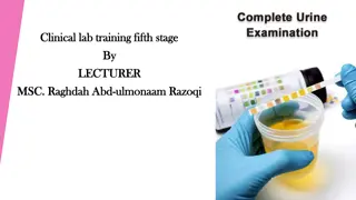
A standard urine test strip may comprise up to 10
different chemical pads or reagents which react (change
color) when immersed in, and then removed from, a urine
sample. The test can often be read in as little as 60 to 120
seconds after dipping.
Routine testing of the urine with multiparameter strips is
the first step in the diagnosis of a wide range of diseases.
The analysis includes testing for the presence
of proteins, glucose, ketones, haemoglobin, bilirubin, uro
bilinogen, acetone, nitrite and leucocytes as well as
testing of pH and specific gravity or to test for infection
by different pathogens.


C
h
e
m
i
c
a
l
 
A
n
a
l
y
s
i
s
 
 
M
i
c
r
o
s
c
o
p
i
c
 
E
x
a
m
i
n
a
t
i
o
n
 
In microscopy, a sample of urine is centrifuged to obtain
some sediment, a
 variety of normal and abnormal cellular
elements may be seen in urine when looked at under a
microscope, including:
Red blood cells
White blood cells
Epithelial cells
Crystals
Bacteria
 
M
i
c
r
o
s
c
o
p
i
c
 
E
x
a
m
i
n
a
t
i
o
n
 
Red Blood Cells:
Hematuria
 is the presence of abnormal numbers of red cells in urine due
to:
a. Glomerular damage
b. Tumors
c. Urinary tract stones
d. Upper and lower urinary tract infections
 
M
i
c
r
o
s
c
o
p
i
c
 
E
x
a
m
i
n
a
t
i
o
n
 
Two Types of Hematuria
Gross hematuria
 
means that the blood can be seen by the naked eye. The
urine may look pinkish, brownish, or bright red.


Microscopic hematuria
 means that the urine is clear, but blood cells can be
seen under a microscope.
 
M
i
c
r
o
s
c
o
p
i
c
 
E
x
a
m
i
n
a
t
i
o
n
 
White blood cells: 
Pyuria
 refers to the presence of abnormal numbers of
leukocytes that may appear with infection in either the upper or
lower urinary tract or with acute glomerulonephritis.
    WBCs - ≤2-5 WBCs/hpf
 
M
i
c
r
o
s
c
o
p
i
c
 
E
x
a
m
i
n
a
t
i
o
n
 
Epithelial cells: 
Renal tubular epithelial cells, contain a
large round or oval nucleus and normally slough into the
urine in small numbers. However, with 
nephrotic
syndrome 
and in conditions leading to 
tubular
degeneration, the number sloughed is increased.
Normal range: ≤15-20 squamous epithelial cells/hpf
 
 
M
i
c
r
o
s
c
o
p
i
c
 
E
x
a
m
i
n
a
t
i
o
n
 
Urinary casts may be made up of cells (such as white
blood cells, red blood cells, kidney cells) or substances
such as protein.
Hyaline casts 
are composed primarily of a mucoprotein
(
Tamm-Horsfall proteins
) secreted by tubule cells.
 
 
M
i
c
r
o
s
c
o
p
i
c
 
E
x
a
m
i
n
a
t
i
o
n
 
Bence Jones proteins are small proteins found in the urine.
Testing for these proteins is done to diagnose and monitor
multiple myeloma 
and other similar diseases.
Bence Jones proteins are considered the 
first tumor marker.
 
A 
tumor marker 
is a substance, made by the body, that is
linked to a certain cancer, or malignancy. Bence Jones
proteins (myeloma casts) are made by plasma cells, a type
of white blood cell. The presence of these proteins in a
person's urine is associated with a malignancy of plasma
cells.


G
e
n
e
r
a
l
 
S
t
o
o
l
 
E
x
a
m
i
n
a
t
i
o
n
 
 
 A stool analysis is a test on a stool (feces) sample to help
diagnose certain conditions affecting the digestive tract.
Collection of Fecal Specimens:
Collect about 10-15 gm the stool in a dry, clean, container.
Make sure no urine, water, soil or other material gets in the
container.
If it is not possible to obtain feces collect a specimen by
inserting a cotton wool swab into rectum for about 10 sec.
Label the specimen with the patient’s name, date and time
of collection and send it with a request form to reach
laboratory as soon as possible.
Fresh stool should be examined immediately, or preserved .
 
G
e
n
e
r
a
l
 
S
t
o
o
l
 
E
x
a
m
i
n
a
t
i
o
n
 
Direct saline wet mount
Place a drop of saline on the slide.
Pick up a small amount of fecal material on the end of an
applicator stick.
Emulsify in the saline and cover with a cover slip.
Examine on low and high power.
The entire preparation must be examined for the
presence of eggs, larvae and protozoa.
Note: 
Take small amounts of material from several
different areas (stool surface and deep inside), especially
from bloody and/or mucoid areas.
 
S
t
o
o
l
 
a
n
a
l
y
s
i
s
 
B
a
s
i
c
s
 
Stool analysis consists of the following:
Macroscopic (Physical) Examination
Chemical Analysis
Microscopic Examination
 
Macroscopic (Physical) Examination
 
1- The color of stool
2- Odor
3- 
Consistency of stool
4-
 Naked eye parasite
5-
 Gross blood
6- 
Mucus


M
a
c
r
o
s
c
o
p
i
c
 
(
P
h
y
s
i
c
a
l
)
 
E
x
a
m
i
n
a
t
i
o
n
 
Normal color 
of stool 
is 
brown
. 
The characteristic brown color of
feces is due to 
stercobilin 
and 
urobinin
, both of which are produced
by bacterial degradation of bilirubin.
Abnormal color:
-
Black color: 
indicate iron medication (for treatment of anemia ) or
upper GIT bleeding (due to peptic ulcer or stomach carcinoma).
-
Bright red color (Hematochezia): 
indicate lower GIT bleeding (due to
anal fissure).
-
Pale brown color: 
with a greasy consistency indicate pancreatic
deficiency causing malabsorption of fat (often with offensive odor).
-
Yellow-green color: 
occurs in the stool of breast-fed infants who lack
normal intestinal flora (low bile conversion) and may also occurs due
to rapid transit of feces through the intestines.
-
Red brown color: 
indicate drugs as Tetracyclines, and Rifambicin
antibiotics.
 
M
a
c
r
o
s
c
o
p
i
c
 
(
P
h
y
s
i
c
a
l
)
 
E
x
a
m
i
n
a
t
i
o
n
 
Normal odor of stool is offensive results from gases produced by
bacterial metabolism, including skatole, mercaptans, indole and
hydrogen sulfide formed by bacterial fermentation and putrefaction.
-
Abnormal odor is 
Very offensive: 
usually seen in cases of constipation
and with certain types of food that produce excessive gases, Bacterial
infection and malabsorption.
Consistency: 
Normally well formed
Abnormal:
-
Very hard: 
seen in cases of constipation
-
Semi formed: 
seen in the cases of parasitic infection
-
Soft: 
seen in the cases of parasitic infection
-
Loose: 
seen in the cases of diarrhea
-
Watery: 
mostly seen in cases of bacterial infection


M
a
c
r
o
s
c
o
p
i
c
 
(
P
h
y
s
i
c
a
l
)
 
E
x
a
m
i
n
a
t
i
o
n
 
Naked eye parasite : Normal: 
no parasites or larva appear in the stool but in
some cause the whole worm or part of its body appear in the stool and can be
seen by naked eye like(segment of tap worm )
-
Two worms can be seen by naked eye in the stool:
-
Ascaris lumbericoides
 
and 
Entrobius vermicularis


Gross blood: Normally no blood seen in the stool.
-
Abnormal 
fresh blood (
Hematochezia
) seen in cases of lower GIT bleeding
and also known as "bright red blood per rectum" and abbreviated 
BRBPR
 
M
a
c
r
o
s
c
o
p
i
c
 
(
P
h
y
s
i
c
a
l
)
 
E
x
a
m
i
n
a
t
i
o
n
 
Mucus: Normally undetectable amount produce by GIT and found in
the stool.
-
Abnormal mucus in the sample appears as white patches and
according to the amount of mucus it can be graded using signs (+,
++, +++).
 
Mucus abnormally can be found in the stool in the following cases:
Spastic colon (translucent mucus on the surface of stool)
Ulcerative colitis
Bacillary dysentery (mucus with fresh pus)
Amoebic dysentery (mucus with fresh blood)
 
C
h
e
m
i
c
a
l
 
A
n
a
l
y
s
i
s
 
pH is Normally variable and diet dependent and is based on
bacterial fermentation in the small intestine.
-
High alkaline stool- Physiological: cause by using High protein
diet
-
 Pathological : Colitis or Antibiotic use (impaired colonic
fermentation)
-
High acidic stool- Physiological :High carbohydrate diet
-
Pathological: Poor fat absorption
 
Fat in stool: Increased Fats is associated with Malabsorption
Syndromes, such as Coeliac disease, Crohn’s disease, Enteritis
and Pancreatic diseases, Surgical removal of section of
Intestine, and others.
 
 
C
h
e
m
i
c
a
l
 
A
n
a
l
y
s
i
s
 
Faecal occult blood 
test (FOBT): 
Detect blood which is
present in amount or form not visible macroscopically.
When the bleeding is chronic with only small amounts of blood
being passed in the feces, the blood (or its breakdown
products) is not recognized in the feces and is referred to as
occult (hidden) blood.
An average, healthy person passes up to 2.0 mL of blood per
150 g of stool into the GIT daily. Passage of more than 2.0
mL of blood in the stool in 24 hours is pathologically
significant.
 
C
h
e
m
i
c
a
l
 
A
n
a
l
y
s
i
s
 
BENZIDINE TEST, GUAIAC TEST, and ORTHOTOLIDINE TEST are used in
FOBT
Principle of the test: The principle of chemical tests to detect occult
blood is based on the fact that hemoglobin and its derivatives react in
a similar way to peroxidase enzymes– by catalyzing the transfer of an
oxygen atom from the peroxide to a chromogen such as benzidine, o-
toludine, guaiac or aminophenazone. Oxidation of the chromogen is
indicated by the production of a blue, blue-green or pink color.


M
i
c
r
o
s
c
o
p
i
c
 
E
x
a
m
i
n
a
t
i
o
n
 
The microscopic examination may be used to determine the presence of
leukocytes and erythrocytes in a fecal smear. This will very quickly give the
clinician information on the patients disease state. Determination of the
presence of leukocytes may be useful in the diagnosis of patients with
diarrhea due to bacterial infection.
Microscopic examination of stool can be done by 
Gram stain
, 
Wrights Stain,
and by wet mount procedure.
 
M
i
c
r
o
s
c
o
p
i
c
 
E
x
a
m
i
n
a
t
i
o
n
 
 
For the diagnosis of microscopic elements:
 
Trophozoites 
and its movements are better seen in
unstained 
preparation of a fresh material.
Cystic forms &Nuclear character 
are better seen in
stained 
preparation(iodine)
Gycogen mass- stained with iodine
Chromatoid bars- unstained preparation
N.B – Both stained and unstained materials are
to be prepared
 
M
i
c
r
o
s
c
o
p
i
c
 
E
x
a
m
i
n
a
t
i
o
n
 
Procedure for the microscopic examination of faecal samples for parasites
1. place a drop of saline a clean slide.
2. place a small piece of stool on the slide and mix with saline, cover with a
cover slip. If the specimen contain mucus, the examination prefer to be done
without saline. The mucus is put on the slide and covered with cover slip.
3. examine under 10X and 40X objectives.
 
Using of Saline: Normal saline (0.85%) is used for routine examination of stool
samples, as it is isotonic.
Using of Iodine: Iodine is used to examine the nuclei of cysts.
Using of Eosin 1%: this provide a pink background and that will help to clear
the unstained objects.
 
T
T
h
h
a
a
n
n
k
k
 
 
Y
Y
o
o
u
u
 
R
e
f
e
r
e
n
c
e
s
 
Urinalysis: Part 1. 2006 The University of Iowa (accessed 17 Aug 2007)
Available from:
http://www.medicine.uiowa.edu/cme/clia/modules.asp?testID=19
http://www.utmem.edu/nephrology/documents/powerpoint-urinalysis-
files/frame.htm
http://www.texascollaborative.org/spencer_urinalysis/ds_overview.htm
http://www.nlm.nih.gov/medlineplus/ency/article/003583.htm
http://www.nlm.nih.gov/medlineplus/ency/article/003587.htm
http://www.medicineplus.com


Slide Note
Embed
Share

General urine examination involves analyzing the color, clarity, and chemical composition of urine, providing insights into various health conditions and potential issues. It helps in detecting metabolic disorders, kidney diseases, and drug abuse. Additionally, the general stool examination is essential for evaluating digestive health. Both examinations are crucial diagnostic tools used by healthcare professionals to assess a patient's overall well-being.


Uploaded on Aug 01, 2024 | 0 Views


Download Presentation

Please find below an Image/Link to download the presentation.

The content on the website is provided AS IS for your information and personal use only. It may not be sold, licensed, or shared on other websites without obtaining consent from the author. Download presentation by click this link. If you encounter any issues during the download, it is possible that the publisher has removed the file from their server.

E N D

Presentation Transcript


  1. General Urine Examination and General Stool Examination Asst. Prof. Dr. Dalya Basil Hanna

  2. Urine analysis Urine is a liquid mainly consisting of water and waste products of our body, generated through filtering the blood by the kidneys. Urine contains residual nitrogen substances such as urea, uric acid, creatinine and a small dose of mineral salts and enzymes. Among these the urochrome, which is the pigment that gives urine the typical amber yellow color. Bilirubin and urobilin also make the urine yellow. substances, there is also

  3. Urine analysis Urine analysis: is the term used to refer to the test used to evaluate a urine sample. Because urine removes liquids from the body, its contents can provide vital health information. Obvious abnormalities in the color, clarity, and cloudiness may suggest different diseases, such as metabolic disorders and kidney disease. Urinalysis can also be used to uncover evidence of drug abuse. toxins and excess

  4. Urinalysis Basics Urinalysis consists of the following: Macroscopic Examination Chemical Analysis Microscopic Examination

  5. Macroscopic Examination Observing the physical properties of the urine: Color: Normal urine should be a shade of yellow ranging from a straw to amber color. Normal urine volume is 750 to 2000 ml/24hr. Abnormal urine can be: colorless, dark yellow, orange, pink, red, green, brown, or black. Clarity (transparency): Normal urine should be clear Abnormal urine can be: hazy, cloudy, or turbid

  6. Macroscopic Examination Pale straw color: Normal, healthy, well-hydrated. Transparent: Colorless urine may indicate over-hydration which can dilute essential salts, such as electrolytes, creating a chemical imbalance in the blood. Transparent yellow: Normal. Dark yellow: Normal, but suggestive of mild dehydration. Amber or honey: Possibly dehydrated. Light orange: Possibly dehydrated, but may also be caused by liver or bile duct problems. Orange: Some medications, such as rifampin or phenazopyridine, can cause this coloration.

  7. Macroscopic Examination Red: This color could be a worrisome sign of many things. Blood in the urine, called hematuria, idiopathic or a sign of a kidney stone, infection or tumor in the urinary tract. It may signal a problem with the prostate. Or a group of as porphyrias. rare inherited disorders known Blue: Some medications and food dyes produce bluish urine. So too does a rare inherited metabolic hypercalcemi or blue diaper syndrome, which is characterized by incomplete intestinal breakdown of tryptophan, a dietary nutrient. disorder known as familial Dark brown or black: Some medications darken urine. Copper or phenol poisoning or melanoma, can result in blackish urine called melanuria. White or milky: This may be caused by an overabundance of certain minerals, such as calcium or phosphate, a urinary tract infection or excessive proteins.

  8. Chemical Analysis The chemical properties of urine, including pH, specific gravity, protein content, glucose content, ketone content, and others are tested. pH Test measures if urine is acidic, basic or neutral Normal urine ranges from 4.6 to 8.0 Specific Gravity: Test measures the concentration of particles in the urine and evaluates the body s water balance. The more concentrated the urine, the higher the urine specific gravity. The most common increase in urine specific gravity is the result of dehydration. Normal urine ranges between 1.002 to 1.028

  9. Chemical Analysis Ketones: Test measures the presence or absence of ketones, the endpoint of rapid or excessive fat breakdown, in the urine. Normal urine does not contain ketones Protein: Normal urine levels of proteins (called albumin) are very small, usually approximately 0 to 8 mg/dl. Glucose: The test measures the amount of sugar in a urine sample. Normal urine does not contain glucose.

  10. Chemical Analysis (Urine test strips) A standard urine test strip may comprise up to 10 different chemical pads or reagents which react (change color) when immersed in, and then removed from, a urine sample. The test can often be read in as little as 60 to 120 seconds after dipping. Routine testing of the urine with multiparameter strips is the first step in the diagnosis of a wide range of diseases. The analysis includes of proteins, glucose, ketones, haemoglobin, bilirubin, uro bilinogen, acetone, nitrite and leucocytes as well as testing of pH and specific gravity or to test for infection by different pathogens. testing for the presence

  11. http://1.bp.blogspot.com/-UVhhcgbhfqw/UE4X8rS2v5I/AAAAAAAAB4E/1CFxx2jJ38k/s640/UroTest+Table.pnghttp://1.bp.blogspot.com/-UVhhcgbhfqw/UE4X8rS2v5I/AAAAAAAAB4E/1CFxx2jJ38k/s640/UroTest+Table.png Chemical Analysis

  12. Microscopic Examination In microscopy, a sample of urine is centrifuged to obtain some sediment, a variety of normal and abnormal cellular elements may be seen in urine when looked at under a microscope, including: Red blood cells White blood cells Epithelial cells Crystals Bacteria

  13. Microscopic Examination Red Blood Cells: Hematuria is the presence of abnormal numbers of red cells in urine due to: a. Glomerular damage b. Tumors c. Urinary tract stones d. Upper and lower urinary tract infections

  14. Microscopic Examination Two Types of Hematuria Gross hematuria means that the blood can be seen by the naked eye. The urine may look pinkish, brownish, or bright red. Microscopic hematuria means that the urine is clear, but blood cells can be seen under a microscope.

  15. Microscopic Examination White blood cells: Pyuria refers to the presence of abnormal numbers of leukocytes that may appear with infection in either the upper or lower urinary tract or with acute glomerulonephritis. WBCs - 2-5 WBCs/hpf

  16. Microscopic Examination Epithelial cells: Renal tubular epithelial cells, contain a large round or oval nucleus and normally slough into the urine in small numbers. syndrome and in conditions degeneration, the number sloughed is increased. However, with nephrotic tubular leading to Normal range: 15-20 squamous epithelial cells/hpf

  17. Microscopic Examination Urinary casts may be made up of cells (such as white blood cells, red blood cells, kidney cells) or substances such as protein. Hyaline casts are composed primarily of a mucoprotein (Tamm-Horsfall proteins) secreted by tubule cells.

  18. Microscopic Examination Bence Jones proteins are small proteins found in the urine. Testing for these proteins is done to diagnose and monitor multiple myeloma and other similar diseases. Bence Jones proteins are considered the first tumor marker. A tumor marker is a substance, made by the body, that is linked to a certain cancer, or malignancy. Bence Jones proteins (myeloma casts) are made by plasma cells, a type of white blood cell. The presence of these proteins in a person's urine is associated with a malignancy of plasma cells.

  19. General Stool Examination A stool analysis is a test on a stool (feces) sample to help diagnose certain conditions affecting the digestive tract. Collection of Fecal Specimens: Collect about 10-15 gm the stool in a dry, clean, container. Make sure no urine, water, soil or other material gets in the container. If it is not possible to obtain feces collect a specimen by inserting a cotton wool swab into rectum for about 10 sec. Label the specimen with the patient s name, date and time of collection and send it with a request form to reach laboratory as soon as possible. Fresh stool should be examined immediately, or preserved .

  20. General Stool Examination Direct saline wet mount Place a drop of saline on the slide. Pick up a small amount of fecal material on the end of an applicator stick. Emulsify in the saline and cover with a cover slip. Examine on low and high power. entire preparation presence of eggs, larvae and protozoa. The must be examined for the Note: Take small amounts of material from several different areas (stool surface and deep inside), especially from bloody and/or mucoid areas.

  21. Stool analysis Basics Stool analysis consists of the following: Macroscopic (Physical) Examination Chemical Analysis Microscopic Examination

  22. Macroscopic (Physical) Examination 1- The color of stool 2- Odor 3- Consistency of stool 4- Naked eye parasite 5- Gross blood 6- Mucus

  23. Macroscopic (Physical) Examination Normal color of stool is brown. The characteristic brown color of feces is due to stercobilin and urobinin, both of which are produced by bacterial degradation of bilirubin. Abnormal color: Black color: indicate iron medication (for treatment of anemia ) or upper GIT bleeding (due to peptic ulcer or stomach carcinoma). - Bright red color (Hematochezia): indicate lower GIT bleeding (due to anal fissure). - Pale brown color: with a greasy consistency indicate pancreatic deficiency causing malabsorption of fat (often with offensive odor). - Yellow-green color: occurs in the stool of breast-fed infants who lack normal intestinal flora (low bile conversion) and may also occurs due to rapid transit of feces through the intestines. - Red brown color: indicate drugs as Tetracyclines, and Rifambicin antibiotics. -

  24. Macroscopic (Physical) Examination Normal odor of stool is offensive results from gases produced by bacterial metabolism, including skatole, mercaptans, indole and hydrogen sulfide formed by bacterial fermentation and putrefaction. Abnormal odor is Very offensive: usually seen in cases of constipation and with certain types of food that produce excessive gases, Bacterial infection and malabsorption. - Consistency: Normally well formed Abnormal: Very hard: seen in cases of constipation - Semi formed: seen in the cases of parasitic infection - Soft: seen in the cases of parasitic infection - Loose: seen in the cases of diarrhea - Watery: mostly seen in cases of bacterial infection -

  25. Macroscopic (Physical) Examination Naked eye parasite : Normal: no parasites or larva appear in the stool but in some cause the whole worm or part of its body appear in the stool and can be seen by naked eye like(segment of tap worm ) Two worms can be seen by naked eye in the stool: - Ascaris lumbericoides and Entrobius vermicularis - Gross blood: Normally no blood seen in the stool. Abnormal fresh blood (Hematochezia) seen in cases of lower GIT bleeding and also known as "bright red blood per rectum" and abbreviated BRBPR -

  26. Macroscopic (Physical) Examination Mucus: Normally undetectable amount produce by GIT and found in the stool. Abnormal mucus in the sample appears as white patches and according to the amount of mucus it can be graded using signs (+, ++, +++). - Mucus abnormally can be found in the stool in the following cases: Spastic colon (translucent mucus on the surface of stool) Ulcerative colitis Bacillary dysentery (mucus with fresh pus) Amoebic dysentery (mucus with fresh blood)

  27. Chemical Analysis pH is Normally variable and diet dependent and is based on bacterial fermentation in the small intestine. High alkaline stool- Physiological: cause by using High protein diet Pathological : Colitis or Antibiotic use (impaired colonic fermentation) High acidic stool- Physiological :High carbohydrate diet Pathological: Poor fat absorption - - - - Fat in stool: Increased Fats is associated with Malabsorption Syndromes, such as Coeliac disease, Crohn s disease, Enteritis and Pancreatic diseases, Surgical Intestine, and others. removal of section of

  28. Chemical Analysis Faecal occult blood test (FOBT): Detect blood which is present in amount or form not visible macroscopically. When the bleeding is chronic with only small amounts of blood being passed in the feces, the blood (or its breakdown products) is not recognized in the feces and is referred to as occult (hidden) blood. An average, healthy person passes up to 2.0 mL of blood per 150 g of stool into the GIT daily. Passage of more than 2.0 mL of blood in the stool in 24 hours is pathologically significant.

  29. Chemical Analysis BENZIDINE TEST, GUAIAC TEST, and ORTHOTOLIDINE TEST are used in FOBT Principle of the test: The principle of chemical tests to detect occult blood is based on the fact that hemoglobin and its derivatives react in a similar way to peroxidase enzymes by catalyzing the transfer of an oxygen atom from the peroxide to a chromogen such as benzidine, o- toludine, guaiac or aminophenazone. Oxidation of the chromogen is indicated by the production of a blue, blue-green or pink color.

  30. Microscopic Examination The microscopic examination may be used to determine the presence of leukocytes and erythrocytes in a fecal smear. This will very quickly give the clinician information on the patients disease state. Determination of the presence of leukocytes may be useful in the diagnosis of patients with diarrhea due to bacterial infection. Microscopic examination of stool can be done by Gram stain, Wrights Stain, and by wet mount procedure.

  31. Microscopic Examination For the diagnosis of microscopic elements: Trophozoites and its movements are better seen in unstained preparation of a fresh material. Cystic forms &Nuclear character are better seen in stained preparation(iodine) Gycogen mass- stained with iodine Chromatoid bars- unstained preparation N.B Both stained and unstained materials are to be prepared

  32. Microscopic Examination Procedure for the microscopic examination of faecal samples for parasites 1. place a drop of saline a clean slide. 2. place a small piece of stool on the slide and mix with saline, cover with a cover slip. If the specimen contain mucus, the examination prefer to be done without saline. The mucus is put on the slide and covered with cover slip. 3. examine under 10X and 40X objectives. Using of Saline: Normal saline (0.85%) is used for routine examination of stool samples, as it is isotonic. Using of Iodine: Iodine is used to examine the nuclei of cysts. Using of Eosin 1%: this provide a pink background and that will help to clear the unstained objects.

  33. Thank You

  34. References Urinalysis: Part 1. 2006 The University of Iowa (accessed 17 Aug 2007) Available from: http://www.medicine.uiowa.edu/cme/clia/modules.asp?testID=19 http://www.utmem.edu/nephrology/documents/powerpoint-urinalysis- files/frame.htm http://www.texascollaborative.org/spencer_urinalysis/ds_overview.htm http://www.nlm.nih.gov/medlineplus/ency/article/003583.htm http://www.nlm.nih.gov/medlineplus/ency/article/003587.htm http://www.medicineplus.com
Related


More Related Content

giItT1WQy@!-/#giItT1WQy@!-/#giItT1WQy@!-/#giItT1WQy@!-/#giItT1WQy@!-/#