Comprehensive Overview of the Nervous System

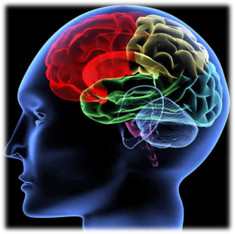
Nervous System
Nervous System
 undefined
OBJECTIVES
OBJECTIVES
At the end of the lecture, students should be able to:
List the subdivisions of the nervous system.
Define the terms: grey matter, white matter, nucleus, ganglion, tract and
nerve.
List the parts of the brain.
Identify the external and internal features of spinal cord.
Enumerate the cranial nerves
Describe the parts and distribution of the spinal nerve.
Define the term ‘dermatome’
List the structures protecting the central nervous system
FUNCTIONS
FUNCTIONS
The nervous system has three
functions:
Collection of Sensory Input
:
Identifies changes occurring
inside and outside the body by
using sensory receptors. These
changes are called stimuli
Integration
:
Processes, analyses and
interprets these changes and
makes decisions
Motor Output:
It then effects a response by
activating muscles or glands
(effectors) via motor output
ORGANIZATION
ORGANIZATION
STRUCTURAL
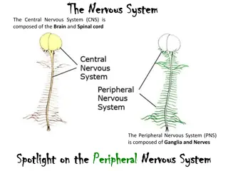
Central Nervous System (CNS)
Brain & Spinal Cord
Peripheral Nervous System
(PNS)
Nerves & Ganglia
ORGANIZATION
ORGANIZATION
FUNCTIONAL
Sensory Division (Afferent)
Motor Division (Efferent)
Autonomic
Somatic
NERVOUS TISSUE
NERVOUS TISSUE
Nervous tissue is
organized as:
Grey matter:
which contains the
cell bodies & the processes of
the neurons, the neuroglia and
the blood vessels.
White matter:
which contains
the processes of the neurons
(no cell bodies), the neuroglia
and the blood vessels.
Nucleus
A group of neurons
within the CNS
Ganglion
A group of neurons
outside the CNS
Tract
A group of nerve fibers
(axons) within the CNS
Nerve
A group of nerve fibers
(axons) outside the CNS
Large mass of nervous tissue located in the cranial cavity.
Has four major regions.
Cerebrum
(Cerebral hemispheres)
Cerebellum
Brainstem:
Midbrain, Pons & Medulla oblongata
Diencephalon:
Thalamus,
Hypothalamus,
Subthalamus &
Epithalamus
THE BRAIN
THE BRAIN
CEREBRUM
CEREBRUM
The largest part of the brain, and has
two hemispheres.
The cerebral hemispheres are
connected by a thick bundle of nerve
fibers called
corpus callosum.
The surface shows ridges of tissue,
called
gyri
,
separated by grooves
called
sulci.
Divided into
4
lobes
by deeper
grooves.
Tissue of Cerebral Hemispheres
Tissue of Cerebral Hemispheres
The outermost layer is called gray matter or cortex.
Deeper is located the white matter, composed of fiber tracts (bundles of
nerve fibers)
Carrying impulses to and from the cortex.
Located deep within the white matter are masses of grey matter called the
basal nuclei .
They help the motor cortex in the regulation of voluntary motor activities
CEREBLLUM
CEREBLLUM
T
h
e
c
e
r
e
b
e
l
l
u
m
h
a
s
2
h
e
m
i
s
p
h
e
r
e
s
a
n
d
a
c
o
n
v
o
l
u
t
e
d
s
u
r
f
a
c
e
.
It has an outer cortex made from gray matter and an inner region of white
matter.
It provides precise coordination for body movements and helps maintain
equilibrium.
QUESTION
QUESTION
4 REVIEW!
4 REVIEW!
Whish statement of the following is NOT true?
Cerebrum has five major regions.
White matter contains the cell bodies & the processes of the neurons, the
neuroglia and the blood vessels.
The innermost layer of cerebrum is called gray matter or cortex.
Cerebellum provides precise coordination for body movements and helps
maintain equilibrium.
SPINAL CORD
SPINAL CORD
It is a two-way conduction pathway
to the brain & a major reflex center
42-45 cm long, cylindrical in shape,
lies within the vertebral canal.
Extends from
foramen magnum
to
L2 vertebra
Continuous above with medulla
oblongata
Caudal tapering end is called
conus
medullaris
Has 2 enlargements:
cervical
and
lumbosacral
Gives rise to 31 pairs of
spinal
nerves
Group of spinal nerves at the end of
the spinal cord is called
cauda
equina
CROSS SECTION OF SPINAL CORD
CROSS SECTION OF SPINAL CORD
The spinal cord is incompletely
divided into two equal parts,
anteriorly
by a short, shallow
median fissure and
posteriorly
by a deep narrow septum, the
posterior median septum.
Composed of grey matter in the
center surrounded by white
matter
The arrangement of grey
matter resembles the shape of
the letter H, having two
posterior, two anterior and two
lateral horns/columns.
PEREPHERAL NERVES
PEREPHERAL NERVES
Cranial
:
12 pairs,
attached to brain,
named, and numbered
from1-12
May be sensory, motor or mixed
Two types:
Spinal
:
31 pairs, attached to
spinal cord named and
numbered according to the
region of the spinal cord
CRANIAL NERVES
CRANIAL NERVES
12 pairs
4 pairs are mixed
trigeminal nerve. (5th)
facial nerve. (7th)
glossopharyngeal nerve. (9th)
vagus nerve. (10th)
5 pairs are motor
occulomotor nerve. (3rd)
trochlear nerve. (4th)
abducent nerve. (6th)
accessory nerve. (11th)
hypoglossal nerve. (12th)
3 pairs are sensory
olfactory nerve. (1st)
optic nerve. (2nd)
vestibulocochlear nerve. (8th)
SPINAL NERVES & NERVE PLEXUES
SPINAL NERVES & NERVE PLEXUES
31 pairs
Each spinal nerve is attached
by two roots:
D
orsal (sensory)
Ventral (motor)
Dorsal root bears a
sensory ganglion
Each spinal nerve exits from
the intervertebral foramen
and divides into a dorsal and
ventral ramus
The rami contain both sensory
and motor fibers
SPINAL NERVES & NERVE PLEXUES
SPINAL NERVES & NERVE PLEXUES
The
dorsal rami
are
distributed individually.
Supply the skin and
muscles of the back
the
ventral rami
form
plexuses
(except in thoracic
region where they form the
intercostal nerves)
Supply the anterior part
of the body
DERMATOME
DERMATOME
Dermatome is a
segment of skin
supplied by one
spinal nerve.
PROTECTION OF CNS
PROTECTION OF CNS
THE CNS IS PROTECTED BY:
Skull
and the
vertebral column
(bone)
Meninges
(membranes): 3 layers
dura mater
(outermost)
arachnoid mater
(middle
pia mater
(innermost)
Cerebrospinal fluid
in the subarachnoid space
CEREBRAL FLUID
CEREBRAL FLUID
Most of the CSF drains from
the ventricles into the
subarachoid space around
the brain and spinal cord. A
little amount flows down in
the central canal of the
spinal cord.
CSF is constantly drained
into the dural sinuses
through the arachnoid villi.
QUESTION
QUESTION
4 REVIEW!
4 REVIEW!
Whish statement of the following is true?
Spinal Cord extends from foramen magnum to L2 vertebra
Dermatome is a segment of skin supplied by one spinal nerve.
CSF is constantly produced by the choroid plexuses outside the ventricles of
brain.
Spinal Cord is incompletely divided into two equal parts, anteriorly by median
fissure and posteriorly by posterior median septum.
Questions!
Questions!
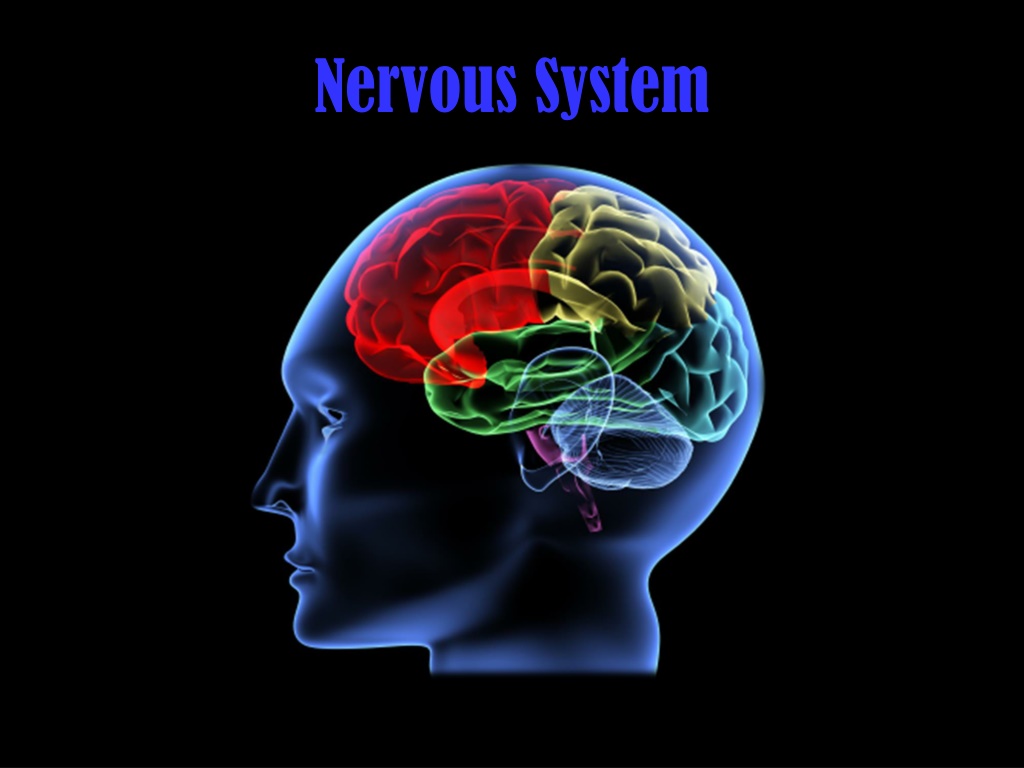
Explore the anatomy and functions of the nervous system with details on its subdivisions, brain parts, spinal cord features, cranial nerves, and more. Learn about sensory input collection, integration, and motor output functions. Understand the structural and functional organization of the central and peripheral nervous systems. Discover the composition of nervous tissue, the roles of grey and white matter, and definitions of key terms like nucleus, ganglion, tract, and nerve. Dive into the complexities of the brain's regions, including the cerebrum, diencephalon, cerebellum, and brainstem.

Download Presentation

Please find below an Image/Link to download the presentation.
The content on the website is provided AS IS for your information and personal use only. It may not be sold, licensed, or shared on other websites without obtaining consent from the author. Download presentation by click this link. If you encounter any issues during the download, it is possible that the publisher has removed the file from their server.
E N D
Presentation Transcript
OBJECTIVES At the end of the lecture, students should be able to: List the subdivisions of the nervous system. Define the terms: grey matter, white matter, nucleus, ganglion, tract and nerve. List the parts of the brain. Identify the external and internal features of spinal cord. Enumerate the cranial nerves Describe the parts and distribution of the spinal nerve. Define the term dermatome List the structures protecting the central nervous system
FUNCTIONS The nervous system has three functions: Collection of Sensory Input: Identifies changes occurring inside and outside the body by using sensory receptors. These changes are called stimuli Integration: Processes, analyses and interprets these changes and makes decisions Motor Output: It then effects a response by activating muscles or glands (effectors) via motor output
ORGANIZATION STRUCTURAL Central Nervous System (CNS) Brain & Spinal Cord Peripheral Nervous System (PNS) Nerves & Ganglia
ORGANIZATION FUNCTIONAL Sensory Division (Afferent) Motor Division (Efferent) Autonomic Somatic
NERVOUS TISSUE Nervous tissue is organized as: Grey matter: which contains the cell bodies & the processes of the neurons, the neuroglia and the blood vessels. White matter: which contains the processes of the neurons (no cell bodies), the neuroglia and the blood vessels.
Nucleus Ganglion A group of neurons within the CNS A group of neurons outside the CNS Nerve Tract A group of nerve fibers (axons) outside the CNS A group of nerve fibers (axons) within the CNS
THE BRAIN Large mass of nervous tissue located in the cranial cavity. Has four major regions. Cerebrum (Cerebral hemispheres) Diencephalon: Thalamus, Hypothalamus, Subthalamus & Epithalamus Cerebellum Brainstem: Midbrain, Pons & Medulla oblongata
CEREBRUM The largest part of the brain, and has two hemispheres. The cerebral hemispheres are connected by a thick bundle of nerve fibers called corpus callosum. The surface shows ridges of tissue, called gyri, separated by grooves called sulci. Divided into 4 lobes by deeper grooves.
Tissue of Cerebral Hemispheres The outermost layer is called gray matter or cortex. Deeper is located the white matter, composed of fiber tracts (bundles of nerve fibers) Carrying impulses to and from the cortex. Located deep within the white matter are masses of grey matter called the basal nuclei . They help the motor cortex in the regulation of voluntary motor activities
CEREBLLUM The cerebellum has 2 hemispheres and a convoluted surface. It has an outer cortex made from gray matter and an inner region of white matter. It provides precise coordination for body movements and helps maintain equilibrium.
QUESTION 4 REVIEW! Whish statement of the following is NOT true? Cerebrum has five major regions. White matter contains the cell bodies & the processes of the neurons, the neuroglia and the blood vessels. The innermost layer of cerebrum is called gray matter or cortex. Cerebellum provides precise coordination for body movements and helps maintain equilibrium.
SPINAL CORD It is a two-way conduction pathway to the brain & a major reflex center 42-45 cm long, cylindrical in shape, lies within the vertebral canal. Extends from foramen magnum to L2 vertebra Continuous above with medulla oblongata Caudal tapering end is called conus medullaris Has 2 enlargements: cervical and lumbosacral Gives rise to 31 pairs of spinal nerves Group of spinal nerves at the end of the spinal cord is called cauda equina
CROSS SECTION OF SPINAL CORD The spinal cord is incompletely divided into two equal parts, anteriorly by a short, shallow median fissure and posteriorly by a deep narrow septum, the posterior median septum. Composed of grey matter in the center surrounded by white matter The arrangement of grey matter resembles the shape of the letter H, having two posterior, two anterior and two lateral horns/columns.
PEREPHERAL NERVES May be sensory, motor or mixed Two types: Cranial: 12 pairs, attached to brain, named, and numbered from1-12 Spinal: 31 pairs, attached to spinal cord named and numbered according to the region of the spinal cord
CRANIAL NERVES 12 pairs 4 pairs are mixed trigeminal nerve. (5th) facial nerve. (7th) glossopharyngeal nerve. (9th) vagus nerve. (10th) 5 pairs are motor occulomotor nerve. (3rd) trochlear nerve. (4th) abducent nerve. (6th) accessory nerve. (11th) hypoglossal nerve. (12th) 3 pairs are sensory olfactory nerve. (1st) optic nerve. (2nd) vestibulocochlear nerve. (8th)
SPINAL NERVES & NERVE PLEXUES 31 pairs Each spinal nerve is attached by two roots: Dorsal (sensory) Ventral (motor) Dorsal root bears a sensory ganglion Each spinal nerve exits from the intervertebral foramen and divides into a dorsal and ventral ramus The rami contain both sensory and motor fibers
SPINAL NERVES & NERVE PLEXUES The dorsal rami are distributed individually. Supply the skin and muscles of the back the ventral rami form plexuses (except in thoracic region where they form the intercostal nerves) Supply the anterior part of the body
DERMATOME Dermatome is a segment of skin supplied by one spinal nerve.
PROTECTION OF CNS THE CNS IS PROTECTED BY: Skull and the vertebral column (bone) Meninges (membranes): 3 layers dura mater (outermost) arachnoid mater (middle pia mater (innermost) Cerebrospinal fluid in the subarachnoid space
CEREBRAL FLUID CSF is constantly produced by the choroid plexuses inside the ventricles of brain. Most of the CSF drains from the ventricles into the subarachoid space around the brain and spinal cord. A little amount flows down in the central canal of the spinal cord. CSF is constantly drained into the dural sinuses through the arachnoid villi.
QUESTION 4 REVIEW! Whish statement of the following is true? Spinal Cord extends from foramen magnum to L2 vertebra Dermatome is a segment of skin supplied by one spinal nerve. CSF is constantly produced by the choroid plexuses outside the ventricles of brain. Spinal Cord is incompletely divided into two equal parts, anteriorly by median fissure and posteriorly by posterior median septum.