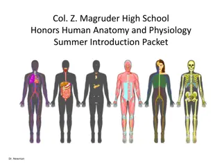
Understanding the Physiology of Postural Reflexes in Maintaining Body Equilibrium
Postural reflexes are automatic movements that help maintain body position and equilibrium during rest or movement. They involve a series of sensory stimuli and motor responses orchestrated by different components of the nervous system. Lesions in these reflex pathways can lead to postural control issues. Exploring the role of the central nervous system, including the spinal cord and brain, in regulating posture is crucial for understanding how the body corrects and adapts to changes in position. Deciphering the intricacies of postural reflexes provides insights into the body's ability to interpret sensory information and make necessary adjustments for maintaining balance.

Download Presentation

Please find below an Image/Link to download the presentation.
The content on the website is provided AS IS for your information and personal use only. It may not be sold, licensed, or shared on other websites without obtaining consent from the author. Download presentation by click this link. If you encounter any issues during the download, it is possible that the publisher has removed the file from their server.
E N D
Presentation Transcript
Physiology of Postural Reflexes Physiology of Postural Reflexes Dr Abdulrahman Alhowikan Collage of medicine Physiology Dep.
Postural reflexes are needed to keep the body in a proper position while standing, moving. Body posture is suddenly altered it is corrected by several reflexes. At spinal cord, medulla, mid- brain and cortical levels. Students are required to know posture regulating parts of cns. Understand these reflexes lesions in the various part of cns To define spinal shock and able to describe the initial and long term changes in spinal reflexes that follow transaction of the spinal cord
Postural reflexes :group of reflexes (automatic movements) which maintain body position and equilibrium either during rest or during movement. It stereotyped motor response to a specific sensory stimulus SENSORY STIMULUS MOTOR RESPONSE E.g Righting reflexes: bring the body into normal position in space and resist forces acting to displace it out of normal position.
Five components 1. Receptor (sensory cell) 2. Sensory neuron 3. Integration center (association neuron, synapses) 4. Motor neuron 5. Effector (muscle or gland cells) 4
play important roles in interpret deeper meanings of the sensory information in the somatosensory areas. it combines information arriving from multiple points in the primary somatosensory area to interpret its meaning. areas 5 and 7, which constitute the somatosensory association area.
If neural axis is transected, the activities integrated below the section are cut off, or released from the control of higher brain centers. Animal experimentation has led to information on the role of cortical and brain stem in control of voluntary movement and posture e.G
Decerebration brain function in an animal by removing the cerebrum, cutting across the brain stem, or severing certain arteries in the brain stem Decerebration is the elimination of cerebral
DECEREBRATION: between the superior and inferior colliculi permits the brain stem pathways to function independent of their input from higher brain structures. This is called a midcollicular and is diagramed in next figure by the dashed line labeled A. DECEREBRATION: transaction of the brain stem midcollicular decerebration decerebration
In midcollicular decerebrate cats, section of dorsal roots to a limb (dashed line labeled B in next figure immediately eliminates the hyperactivity of extensor muscles. This suggests that decerebrate rigidity is spasticity due to facilitation of the myotatic stretch reflex.
If the anterior lobe of the cerebellum is removed in a decerebrate animal (dashed line labeled C in next figure , extensor muscle hyperactivity is exaggerated ( (decerebellate decerebellate rigidity) rigidity)
Removal of the cerebral cortex ( (decortication dashed line labeled D in next figure produces decorticate rigidity flexion of the upper extremities at the elbow and extensor hyperactivity in the lower extremities Decorticate rigidity is seen on the hemiplegic side in humans after hemorrhages or thromboses in the internal capsule. decortication; ; decorticate rigidity which is characterized by
1. Tonic labyrinthic reflexes Receptors: Otolith organs of vestibular apparatus via vestibulospinal tract Stimulus: Gravity, change body position Response: Supine position maximum rigidity in extensor ; Prone position minimum rigidity 2. Tonic neck reflexes Receptors: neck proprioceptors Stimulus: Head turned to side Response: Extension of limbs on side to which head is turned, up-hind leg flex, down-foreleg flex
1. Labyrinthine righting reflexes Receptors: Otholithic organs Stimulus: Gravity Response: Head is kept at level 2. Body on head righting reflexes Stimulus: Pressure on side of body exteroceptors Response: Righting to head (correct head position) 3. Body on body righting reflexes Stimulus: Pressure on side of body exteroceptors Response: Righting of body even when head is prevented to right
4. Body on neck righting reflexes Stimulus: Stretch of neck muscle Response: Contraction of neck muscles rights thorax and abdomen 5. Group reflex When an object is brought close to limbs, animals grasp object and limb are extended. 6. Vestibular placing reaction When blindfolded animal is brought down from height rapidly, forearm of animal extend and toes spread which assist animal to steadily land on ground.
1. Optical righting reflex Stimulus: Visual clues Response: Righting of head 2. Hopping reactions If a standing animal is pushed laterally, it hops (jumps) to maintain equilibrium. 3. Placing reaction Stimulus: Visual, exteroceptive and proprioceptive receptors Response: Foot placed on supporting surface in position to support body
Eliciting the flexor reflex do not pass directly to the anterior motor neurons pass first into the spinal cord interneuron only secondarily to the motor neurons. Pattern of withdrawal Stimulus side of the arm contraction of the flexor muscles and abductor muscles it integrative centers Pattern of withdrawal
following a stimulus elicits a flexor reflex in one limb, the opposite limb begins to extend. signals from sensory nerves cross to the opposite side of the cord to excite extensor muscles the crossed extensor reflex usually does not begin until 200 to 500 milliseconds after onset of the initial pain stimulus,
Excitation of one group of muscles is often associated with inhibition of another group. This is the phenomenon of reciprocal inhibition, and the neuronal circuit called reciprocal innervation It exist between the muscles on the two sides of the body
Positive Supportive Reaction: Pressure on the footpad of a decerebrate animal causes the limb to extend against the pressure applied to the foot magnet reaction: pressure on one side causes extension in that direction ;avoid falling to that side. Cord "Righting" Reflexes: if animal is laid on side uncoordinated movements to be in standing position Rhythmical Stepping Movements of a Single Limb: flexion of limb followed by backward extension. Then flexion occurs again
Reciprocal Stepping of Opposite Limbs: occurs in the forward direction in one limb, the opposite limb ordinarily moves backward. This effect results from reciprocal innervation between the two limbs. Diagonal Stepping of All Four Limbs Reflex stepping occurs diagonally between the forelimbs and hindlimbs. This diagonal response is another manifestation of reciprocal innervation, Reciprocal Stepping of Opposite Limbs: stepping Diagonal Stepping of All Four Limbs Reflex :
Tonic Labyrinthine Reflex https://www.youtube.com/watch?v=75HOIjF x1ME -------------- --------- --------
Suddenly transected of spinal cord in the upper neck, cord reflexes, immediately become depressed to the point of total silence After a few hours to a few weeks, the spinal neurons gradually regain their excitability
Functions affected in spinal shock : arterial blood pressure falls to as low as 40 mm hg Skeletal muscle reflexes are blocked then return to normal; some reflexes become hyperexcitable,. The sacral reflexes for control of bladder and colon evacuation (Constipation) are hidden few weeks after cord transection, but in most cases they eventually return.
Reference book Guyton & Hall: Textbook of Medical Physiology 12E Thank you Thank you