Understanding Arteries: Anatomy and Function Overview
Explore the key concepts related to arteries, including their definition, anastomosis, end arteries, aorta divisions, and major artery distribution in the body. Learn about the general principles of arteries and their role in blood circulation, emphasizing the importance of anastomoses. Dive into the details of the aorta, its structure, and branches. Gain insights into the ascending aorta, arch of aorta, and common carotid artery. Discover the significance of arteries in carrying oxygenated blood and maintaining circulation throughout the body.

Download Presentation

Please find below an Image/Link to download the presentation.
The content on the website is provided AS IS for your information and personal use only. It may not be sold, licensed, or shared on other websites without obtaining consent from the author. Download presentation by click this link. If you encounter any issues during the download, it is possible that the publisher has removed the file from their server.
E N D
Presentation Transcript
OBJECTIVES OBJECTIVES At the end of the lecture, the student should be able to: Define the word artery and understand the general principles of the arterial system. Define arterial anastomosis and describe its significance. Define end arteries and give examples. Describe the aorta and its divisions & list the branches from each part. List major arteries and their distribution in the head & neck, thorax, abdomen and upper & lower extremities. List main pulse points.
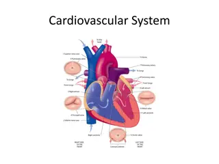
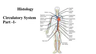
ARTERIES ARTERIES Arteries carry blood from the heart to the body. All arteries, carry oxygenated blood, EXCEPT the PULMONARY ARTERY which carry deoxygenated blood to the lungs.
GENERAL PRINCIPLES OF ARTERIES GENERAL PRINCIPLES OF ARTERIES The flow of blood depends on the pumping action of the heart. Arteries have ELASTIC WALL containing NO VALVES. The branches of arteries supplying adjacent areas normally ANASTOMOSE with one another freely providing backup routes for blood to flow if one artery is blocked, e.g. arteries of limbs. The arteries whose terminal branches do not anastomose with branches of adjacent arteries are called END ARTERIES . End arteries are of two types: Anatomic (True) End Artery: When NO anastomosis exists, e.g. artery of the retina. Functional End Artery: When an anastomosis exists but is incapable of providing a sufficient supply of blood, e.g. splenic artery, renal artery.
AORTA AORTA The largest artery in the body Carries oxygenated blood to all parts of the body Is divided into 4 parts: 1. Ascending aorta 2. Arch of aorta 3. Descending thoracic aorta 4. Abdominal aorta 2 1 3 4
ASCENDING AORTA ASCENDING AORTA Originates from left ventricle. Continues as the arch of aorta Has three dilatations at its base, called aortic sinuses Branches: Right & Left coronary arteries (supplying heart), arise from aortic sinuses
ARCH OF AORTA ARCH OF AORTA Continuation of the ascending aorta. Leads to descending aorta. Located behind the lower part of manubrium sterni and on the left side of trachea. Branches: 1. Brachiocephalic trunk. 2. Left common carotid artery. 3. Left subclavian artery. 12 3
COMMON CAROTID ARTERY COMMON CAROTID ARTERY Origin: LEFT from aortic arch. RIGHT from brachiocephalic trunk. Each common carotid divides into two branches: Internal carotid External carotid
EXTERNAL CAROTID ARTERY EXTERNAL CAROTID ARTERY It divides behind neck of mandible into: Superficial temporal & maxillary arteries It supplies: Scalp: Superficial temporal, occipital, & posterior auricular arteries Face: Facial artery Maxilla & mandible: Maxillary artery Tongue: Lingual artery Pharynx: ascending pharyngeal artery Thyroid gland: Superior thyroid artery 7 8 6 4 5 3 2 1
INTERNAL CAROTID ARTERY INTERNAL CAROTID ARTERY Has NO branches in the neck Enters the cranial cavity, joins the basilar artery (formed by the union of two vertebral arteries) and forms arterial circle of Willis to supply brain. In addition, it supplies Nose Scalp Eye
SUBCLAVIAN ARTERY SUBCLAVIAN ARTERY Origin: LEFT: from arch ofaorta RIGHT: from brachiocephalic trunk It continues, at lateral border of first rib, as axillary artery: artery of upper limb Main branches: Vertebral artery: supplies brain & spinal cord Internal thoracic artery: supplies thoracic wall
ARTERIES OF UPPER LIMB ARTERIES OF UPPER LIMB At lateral border of 1st rib At lower border of teres major Opposite neck of radius
DESCENDING THORACIC AORTA DESCENDING THORACIC AORTA It is the continuation of aortic arch At the level of the 12th thoracic vertebra, it passes through the diaphragm and continues as the abdominal aorta Branches: Pericardial Esophageal Bronchial Posterior intercostal
ABDOMINAL AORTA ABDOMINAL AORTA It enters the abdomen through the aortic opening of diaphragm. At the level of lower border of L4, it divides into two common Iliac arteries. Branches: divided into two groups: Single branches Paired branches
MAIN BRANCHES OF MAIN BRANCHES OF ABDOMINAL AORTA ABDOMINAL AORTA 1 1 2 2 SINGLE BRANCHES SUPPLYING GASTROINTESTINAL TRACT 3 3 4 PAIRED BRANCHES 5
BRANCHES OF COMMON ILIAC ARTERY BRANCHES OF COMMON ILIAC ARTERY EXTERNAL ILIAC ARTERY: continues (at midpoint of inguinal ligament) as femoral artery the main supply for lower limb INTERNAL ILIAC ARTERY: supplies pelvis
ARTERIES OF LOWER LIMB ARTERIES OF LOWER LIMB Femoral Artery Is the main arterial supply to lower limb Is the continuation of external iliac artery behind the midpoint of the inguinal ligament Passes through adductor hiatus and continues as: Popliteal Artery Deeply placed in the popliteal fossa. Divides, at lower end of popliteal fossa into: 1-Anterior Tibial Artery 2-Posterior Tibial Artery
PULSE POINTS IN HEAD & NECK PULSE POINTS IN HEAD & NECK
PULSE POINTS IN UPPER LIMB PULSE POINTS IN UPPER LIMB
PULSE POINTS IN LOWER LIMB PULSE POINTS IN LOWER LIMB
THANK YOU THANK YOU