Understanding Coronary Circulation and Arterial Supply in Cardiovascular Physiology

Explore the intricate details of coronary circulation, including arterial supply, branches of coronary arteries, and areas of distribution. Dr. Abeer Al-Masri, an Associate Professor and Cardiovascular Physiologist at the Faculty of Medicine, KSU, provides valuable insights on the topic, covering the differentiation between right and left coronary arteries, as well as the significance of coronary ostia in delivering oxygenated blood to the cardiac muscle.
- Cardiovascular Physiology
- Coronary Circulation
- Arterial Supply
- Dr. Abeer Al-Masri
- Faculty of Medicine

Download Presentation

Please find below an Image/Link to download the presentation.
The content on the website is provided AS IS for your information and personal use only. It may not be sold, licensed, or shared on other websites without obtaining consent from the author. Download presentation by click this link. If you encounter any issues during the download, it is possible that the publisher has removed the file from their server.
E N D
Presentation Transcript
Dr. Abeer A. Al-Masri Associate Professor & Consultant, Cardiovascular Physiologist, Faculty of Medicine, KSU.
At end of this lecture you should be able to know: 2 Dr. Abeer Al-Masri, Faculty of Medicine, KSU.
Coronary Circulation Consists of: 1. 2. 3. Arterial supply. Venous drainage. Lymphatic drainage. 4 Dr. Abeer Al-Masri, Faculty of Medicine, KSU.
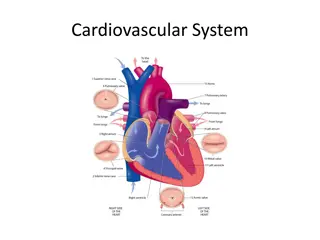
Arterial Supply Cardiac muscle is supplied by two coronary arteries: A. Right coronary artery (RCA.) B. Left coronary artery (LCA.) Both coronaries arise from the coronary sinuses just superior to the aortic valve cusps at the aortic root. 5 Dr. Abeer Al-Masri, Faculty of Medicine, KSU.
I: Arterial Supply The aortic valve has three cusps: left coronary (LC), right coronary (RC) & posterior non-coronary (NC) cusps. Coronary arteries arise just superior to the aortic valvecusps. Coronary Ostia (origins of the coronary arteries) may vary in shape & location, most of which are of no clinical significance. Coronary arteries deliver oxygenated blood to thecardiac muscle. 6 Dr. Abeer Al-Masri, Faculty of Medicine, KSU.
Right vs Left Coronary Arteries Smaller than the leftcoronary artery. Arises from the rightcoronary sinus. Terminates by anastomosing with the left coronaryartery. Larger than the right coronary artery. Arises from the leftcoronary sinus. Terminates by anastomosing with the right coronary artery. 7 Dr. Abeer Al-Masri, Faculty of Medicine, KSU.
Branches of Coronary Arteries Left Coronary Artery (LCA): Right Coronary Artery (RCA): - Right Marginal Artery (RMA) - Left Anterior Descending (LAD) - Posterior Descending Branch - Circumflex Artery (CX) - SA nodal Artery - Left Marginal Artery (LMA) - AV nodal Artery - SA nodal Artery 8 Dr. Abeer Al-Masri, Faculty of Medicine, KSU.
Branches and Areas of Distribution of Coronary Arteries 9 Dr. Abeer Al-Masri, Faculty of Medicine, KSU.
Areas of Distribution of Left Coronary Artery (LCA) LAD supplies: - Anterior & apical parts of the heart: LV & the TVarea adjoining the anterior inter ventricular (IV) groove. - Anterior 2/3rd of the inter ventricular (IV) septum. CX & LMA supplies: - Lateral & posterior surfaces of the heart: LV & SA- node. Dr. Abeer Al-Masri, Faculty of Medicine, KSU. 10
Areas of Distribution of Right Coronary Artery (RCA) RMA supplies: - RV, except the area adjoining the anterior inter ventricular (IV) groove. PDA supplies: - Posterior 1/3rd of the inter ventricular (IV) septum. SA nodal Artery supplies: - The SA- node (Note: in 40% of cases, it is supplied by LCA) - Surrounding Right atrium. - Inferior part of Lt ventricle adjoining the posterior inter ventricular groove. AV nodal Artery supplies: - The AV-node (Note: A part of the Lt branch of AV- Bundle) - Surrounding Right atrium. 11 Dr. Abeer Al-Masri, Faculty of Medicine, KSU.
Areas of Distribution of Coronary Arteries 12 Dr. Abeer Al-Masri, Faculty of Medicine, KSU.
Collateral Circulation Cardiac anastomosis: The two coronary arteries anastomose in the myocardium. Extra cardiac anastomosis: The two coronary arteries anastomosewith: Vasa vasorumoftheaorta. Vasa vasorumofpulmonaryarteries. Internal thoracicarteries. The bronchialarteries. Phrenicarteries. Extra cardiac channels open up in case of emergencies, when the coronary arteries are blocked. 13 Dr. Abeer Al-Masri, Faculty of Medicine, KSU.
II: Venous Drainage of The Heart Venous drainage brings deoxygenated blood back to the heart. Cardiac venous drainage occur through: - Coronary sinus, which lies in the posterior part of the atrioventricular groove & is a continuation of the great cardiac vein. - Anterior, middle & smallcardiac veins. - Venae Cordis Minimae (smallest cardiac veins.) 14 Dr. Abeer Al-Masri, Faculty of Medicine, KSU.
Venous Drainage of The Heart.. Cont. Most of the venous blood return to the heart into the Right atrium through the coronary sinus via the cardiac veins. 5- 10% drains directly into heart chambers, Right atrium & Right ventricle: by the anterior cardiac vein & the small veins that open directly into the heart chambers. 15 Dr. Abeer Al-Masri, Faculty of Medicine, KSU.
III: Lymphatic Drainage of The Heart Lymphatics of the heart accompany the two coronary arteries. Lymphatics of the heart form two trunks: - The right trunk: ends in the brachiocephalic node. - The left trunk: ends into the tracheo-bronchial lymph nodes, at the bifurcation of the trachea. 16 Dr. Abeer Al-Masri, Faculty of Medicine, KSU.
Coronary Dominance Coronary dominance depends on which artery (or arteries) gives rise to the posterior descending artery (PDA), that runs along the posterior side of the heart & supplies the AV- node. A person can be: - Rightdominant, - Left dominant,or - Co-dominant. 17 Dr. Abeer Al-Masri, Faculty of Medicine, KSU.
Coronary Dominance Cont. Coronary dominance is recognized by the presence of septal perforating branches. The right coronary artery is dominant in 80 85% cases. The circumflex branch of the left coronary artery is dominant in 8- 10% cases0 Balanced or co-dominance is found in 7-10% of population where the posterior inter ventricular artery is formed by both Right coronary & LCX arteries. Clinical importance: - In left dominance,a block in LCA affect the entire left ventricle & IV septum. - In right or balanced dominance, a block in RCA at least sparespart (2/3) of the septum & Lt ventricle. 18 Dr. Abeer Al-Masri, Faculty of Medicine, KSU.
Coronary Blood Flow (CBF) Coronary blood flow (CBF) at rest in humans is about 5% of the cardiac output, which is about 225-250 ml/min. CBF increases in proportion to exercise or work output. At rest, the heart extracts 60-70% of oxygen from each unit of blood delivered to the heart, due to the presence of more mitochondria which generate energy for contraction by aerobic metabolism (other tissue extract only 25% of O2.) 19 Dr. Abeer Al-Masri, Faculty of Medicine, KSU.
Phasic Changes in Coronary Blood Flow During Systole & Diastole During systole, coronary arteries are compressed & the blood flow to the left ventricle isreduced. 20 Dr. Abeer Al-Masri, Faculty of Medicine, KSU.
Factors Affecting Coronary Blood Flow (CBF) Pressure gradient across theaorta. Chemical factors. Neural factors. Dr. Abeer Al-Masri, Faculty of Medicine, KSU. 21
Pressure Gradient between Aorta & Ventricles Affects CBF CBF to the Right side is not much affected during systole. Pressure difference between the aorta & Right ventricle is greater during systole than during diastole, therefore more blood flow to Right ventricle occurs during systole. Pressure difference (mmHg) between aorta & Pressure (mmHg) in Left Ventricle Right Ventricle Left Ventricle Right Ventricle Aorta Systole 120 120 25 0 95 Diastole 80 0-2 0-2 80 80 Dr. Abeer Al-Masri, Faculty of Medicine, KSU. 22
Chemical Factors Affecting Coronary Blood Flow Chemical factors causing Coronary vasodilatation (Increased coronary blood flow): Lack of oxygen. Increased local concentration ofCo2. Increased local concentration ofH+ ion. Increased local concentrationof k+ion. Increased local concentration of Lactate, Prostaglandin, Adenosine, Adenine nucleotides. Dr. Abeer Al-Masri, Faculty of Medicine, KSU. 23
Neural Factors Affecting Coronary Blood Flow Sympathetic stimulation. Parasympathetic stimulation. Dr. Abeer Al-Masri, Faculty of Medicine, KSU. 24
Effect of Sympathetic Stimulation on Coronary Blood Flow Coronary arteries have: - Alpha Adrenergic receptors, which mediatevasoconstriction (more epicardial.) - Beta Adrenergic receptors, which mediate vasodilatation (more in the intramusculararteries.) Indirect effect of sympathetic stimulation: Sympathetic stimulation in intact body will lead to release of adrenaline & noradrenaline, increasing HR & forceof contraction. Vasodilator metabolites will be increased leading to coronary vasodilatation. Direct effect of sympathetic stimulation: Experimentally, injection of noradrenalin after blocking of the beta adrenergic receptors in un-anesthetized animalselicits coronary vasoconstriction. Dr. Abeer Al-Masri, Faculty of Medicine, KSU. 25
Benefits of indirect effect of nor- adrenergic discharge When systemic blood pressure decreases very low. Reflex increase of nor adrenergic discharge. Increase CBF secondary to metabolic changes in the myocardium. In this way, circulation of the heart is preserved while the flow to other organs compromised. Dr. Abeer Al-Masri, Faculty of Medicine, KSU. 26
Effect of Parasympathetic Stimulation on Coronary Blood Flow Vagal stimulation (Parasympathetic) causes coronary vasodilatation. However, parasympathetic distribution is not great. There is more sympathetic innervation of coronary vessels. Dr. Abeer Al-Masri, Faculty of Medicine, KSU. 27
Effect of Tachycardia on Coronary Blood Flow CBF is reduced with tachycardia, as the diastolic period will be shortened. Dr. Abeer Al-Masri, Faculty of Medicine, KSU. 28
Control of Coronary Blood Flow (CBF) CBF shows considerable autoregulation. Local muscle metabolism is the primary controller: - Oxygen demand is a major factor in local coronary blood flow regulation. Nervous control of CBF: - Direct effects of nervous stimuli on the coronary vasculature. - Sympathetic greater effects than parasympathetic. Dr. Abeer Al-Masri, Faculty of Medicine, KSU. 29


")