Understanding the Circulatory System and Blood Vessels in Histology

The circulatory system, comprising the cardiovascular and lymphatic systems, plays a vital role in transporting essential substances throughout the body. It consists of arteries, veins, capillaries, and the heart. Blood vessels have distinct histological structures with three main layers - intima, media, and adventitia. Understanding the organization and function of blood vessels is crucial for grasping the dynamics of circulation and maintaining bodily homeostasis.

Download Presentation

Please find below an Image/Link to download the presentation.
The content on the website is provided AS IS for your information and personal use only. It may not be sold, licensed, or shared on other websites without obtaining consent from the author. Download presentation by click this link. If you encounter any issues during the download, it is possible that the publisher has removed the file from their server.
E N D
Presentation Transcript
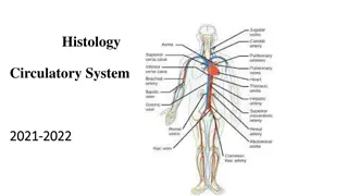
Histology Circulatory System Part -1-
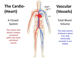
Circulatory System The circulatory system is an organ system that permits blood and lymph circulation to transport nutrients (such as amino acids and electrolytes), oxygen, carbon dioxide, hormones, blood cells, etc. to and from cells in the body to nourish it and help to fight diseases, stabilize body temperature and pH, and to maintain homeostasis. The vascular system is a continuous network of tubes or vessels that distributes blood throughout the body and returns it the heart.
The circulatory system consist of two systems:- 1-Blood vascular system or cardiovascular system. 2-Lymphatic vascular system. A. The cardiovascular system The blood vascular system is composed of the following: 1.Arteries 2. Arterioles 3.Veins 4.Capillaries 5.Heart 6.Venules
B. The lymph vascular system 1. Blind ended lymphatic capillaries that collect lymph fluid from tissues. 2. larger lymphatic vessels that connect with one and other and finally empty collected lymph into large veins in the neck where the lymphatic and cardiovascular systems merge.
General Structure of Blood Vessels Blood vessels of various sizes and should be aware that the histological appearances of vessels of different sizes (arterioles, arteries) and different types (arteries ,veins) are different from each other. These differences are the result of quantitative variations of a common structural pattern that can be seen in all blood vessels with the exception of capillaries, i.e...
the division of the walls of the blood vessels into three layers or tunics . These are arranged into three concentric layers: intima, media and adventitia. The intima is the inner layer abutting the vessel lumen. The adventitia is the outer layer abutting the perivascular soft tissue. The media is sandwiched between the intima and adventitia
1 1 1-The intima is the thinnest layer. It is composed of a single layer of endothelial cells and a small amount of sub endothelial connective tissue. The intima is separated from the media by a dense elastic membrane called the internal elastic lamina. 2-The media is the thickest layer and provides structural support, vasoreactivity and elasticity. It is composed of smooth muscle cells, elastic fibers and connective tissue, which vary in amount depending on the type of vessel. Smooth muscle cells contract (vasoconstriction) or relax (vasodilatation), which is controlled by autonomic nerves (nervivasorum) and local metabolic factors.
3-The adventitia is composed of connective tissue, nutrient vessels (vasa vasorum) and autonomic nerves (nervivasorum). The intima and inner part of the media are nourished by diffusion of oxygen and nutrients from blood in the lumen, and the adventitia and outer part of the media are nourished by vasa vasorum.
Arteries The walls of arteries are thicker than that of veins to withstand pulsatile flow and higher blood pressures. As arteries become smaller, wall thickness gradually decreases but the ratio of wall thickness to lumen diameter increases . Arteries are divided into three types according to size and function. The constituents of the media of these vessels differ in their relative amounts accordingly.
1-Large elastic arteries (aorta, large aortic branchesand pulmonary arteries) the media is abundant in elastic fibers that allow it to expand with systole and recoil during diastole, thereby propelling blood forward. 2-Medium-sized muscular arteries (other aortic branches, coronary and renal arteries) the media is abundant in smooth muscle cells that vasoconstriction or vasodilation, thereby controlling lumen diameter and regional blood flow. 3-Small arteries and arterioles (in the substance of organs and tissues) the media is abundant in smooth muscle cells that vasoconstriction or vasodilation. In vessels of this size, smooth muscle contraction causes dramatic changes in lumen diameter, thereby controlling systemic blood pressure as well as regional blood flow.
Veins The walls of veins are thinner than the walls of arteries, while their diameter is larger. In contrast to arteries, the layering in the wall of veins is not very distinct. The tunica intima is very thin. Only the largest veins contain an appreciable amount of sub endothelial connective tissue. Internal and external elastic laminae are absent or very thin. The tunica media appears thinner than the tunica adventitia, and the two layers tend to blend into each other.
The appearance of the wall of veins also depends on their location. The walls of veins in the lower parts of the body are typically thicker than those of the upper parts of the body, and the walls of veins which are embedded in tissues that may provide some structural support are thinner than the walls of unsupported veins.