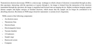
Advanced Techniques in Materials Science: Transmission Electron Microscopy
Explore the advanced techniques used in materials science, focusing on Transmission Electron Microscopy (TEM). Learn about the challenges with optical microscopes, the principles of TEM imaging, and the application of scattering theory in electron microscopy. Discover how TEM offers higher resolutio
3 views • 19 slides
Advanced Microscopy Techniques and Applications in Cell Biology
Explore the diverse world of advanced microscopy techniques in cell biology, from confocal and super-resolution imaging to Fluorescence Resonance Energy Transfer (FRET) microscopy and Fluorescence Recovery After Photobleaching. Discover the latest methods, considerations, and applications for studyi
3 views • 21 slides
Meticulous Research Unveils Comprehensive Insights into the Microscopy Market
Microscopy Market by Product (Microscopes, Software), Type [Optical (Compound, Digital), Electron (SEM, TEM), Scanning Probe (AFM, STM)], Application (Research, Material), and End User (Electronics, Healthcare, F&B, Academic) - Forecast to 2028
7 views • 2 slides
Meticulous Research Releases Comprehensive Analysis of the Microscopy Market
Microscopy Market Size, Share, Forecast, & Trends Analysis by Product (Microscopes (Optical [Compound, Stereo, Digital] Electron [SEM, TEM] Scanning Probe [AFM, STM]), Software) Application (R&D, Material Analysis) End User
0 views • 2 slides
Evolution of Microscopes: From Reading Stones to Phase-Contrast Microscopy
The history of microscopes dates back to the 11th century with the invention of the reading stone, leading to the creation of wearable eyeglasses in the 13th century. The first microscope was developed in the late 16th century by Zacharias Jansen, paving the way for the compound microscope and the t
0 views • 17 slides
Basic Concepts of Microscopy for Materials Study
Microscopy plays a crucial role in examining the structural elements and defects influencing material properties. This involves studying microscopic features like grain size and shape using optical, electron, and scanning probe microscopes. Microstructural examinations help understand property-struc
1 views • 12 slides
Understanding Fluorescence Microscopy: Principles and Applications
Fluorescence microscopy, pioneered by British scientist Sir George G. Stokes, reveals hidden details in specimens using fluorescent dyes that emit light of longer wavelengths. This innovative technique allows for visualization of cellular components that are otherwise colorless under conventional mi
0 views • 19 slides
Understanding Microscopy and Micrometry in Veterinary Microbiology
Microscopy and micrometry play a crucial role in observing small organisms like microorganisms, making them visible for study. This article delves into the history, definition, types of microscopes, variables used in microscopy, and the significance of these tools in veterinary microbiology.
1 views • 15 slides
Principles and Applications of Centrifugation in Microtomy and Microscopy
Centrifugation is a process that separates substances based on density using high-speed rotation. This article explores the basic principles of centrifugation, instrumentation, and applications in the fields of microtomy and microscopy. It covers the different types of centrifuges, such as low-speed
0 views • 28 slides
Understanding Phase Contrast Microscopy: A Revolutionary Tool in Cell Observation
Phase contrast microscopy, invented by Frits Zernike, revolutionized cell observation by utilizing tiny phase changes in light rays to create contrast in living cells. This technique exploits differences in refractive indices to enhance visibility without staining cells, offering a new perspective i
0 views • 27 slides
Analysis and Applications of Electron Microscopy in Science Education
Explore the in-depth analysis of quantitative and qualitative techniques in electron microscopy as it relates to interactions with matter, instrumentation, specimen preparation, elemental analysis, and more. Delve into the electromagnetic spectrum, radiation effects on the human body, and spectrosco
0 views • 13 slides
Bacteria Identification Techniques: Staining and Microscopy Overview
Staining techniques in microbiology play a crucial role in visualizing bacteria under microscopes. This content covers the importance of staining, types of dyes, wet and dry mounts, hanging drop technique, and different types of staining methods employed in identifying bacteria. Explore the basics o
0 views • 39 slides
Exploring Fluorescence Microscopy and High-Speed Cameras in Modern Biology
Delve into the fascinating world of fluorescence microscopy and high-speed cameras in biology through topics such as detectors for microscopy, Nyquist criterion, visualizing hearing in vivo, and temporal resolution insights. Learn about techniques, equipment, and practical considerations in utilizin
1 views • 69 slides
Proper Section Cutting Techniques for Microscopy
Learn about proper section cutting techniques for light and electron microscopy, including the ideal thickness for sections. Discover the essential steps for cutting paraffin sections and common faults to avoid. Find out the key points necessary for easy and successful sectioning, such as equipment
0 views • 4 slides
Understanding The Light Microscope: A Comprehensive Guide
Delve into the world of microscopy with Dr. Mohammed Hussein's Lab Manual No. 1, covering the fundamentals of the light microscope, its components, lenses, magnification power, resolving power, and detailed instructions on how to focus your microscope effectively for optimal results. Explore the his
0 views • 19 slides
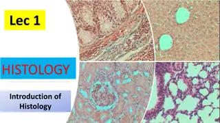
Introduction to Histology and Microscopy in Anatomy
Histology is the study of tissue structure and function in animals and plants, focusing on the microscopic structure of cells, tissues, and organs. It helps in understanding the relationship between structure and function, intertwining disciplines like cell biology, pathology, physiology, and bioche
0 views • 5 slides
Understanding the Compound Light Microscope and Its Applications
Explore the fundamental concepts of microscopy through Professor Diane Hilker's detailed lectures on the use and care of the compound light microscope. Dive into topics like magnification, resolution, and the practical application of microscopes in scientific research. Enhance your understanding of
0 views • 17 slides
Advanced Microscopy Techniques in EUV Lithography: SHARP Overview
SHARP utilizes Fresnel zone plate lenses to achieve diffraction-limited quality in EUV lithography, offering a range of NA values and image magnifications. The system allows emulation of mask-side imaging conditions with hundreds of lenses available. Coherence control and engineering are provided th
1 views • 18 slides
Next-Generation Photomasks in Extreme Ultraviolet Lithography Symposium
Explore the latest advancements in actinic mask imaging and semiconductor high-NA reticle technology presented at the 2015 International Symposium on Extreme Ultraviolet Lithography in Maastricht. Topics include zoneplate lenses, phase reconstruction algorithms, Fourier Ptychography microscopy, and
0 views • 56 slides
Laboratory Diagnosis of Malaria: Methods and Procedures
Methods for diagnosing malaria in laboratory settings involve techniques such as light microscopy and rapid diagnostic tests. Light microscopy is used to prepare blood films, enabling the detection of malaria parasites like Plasmodium falciparum. Rapid diagnostic tests can quickly identify malaria a
0 views • 26 slides
Understanding Cell Structure and Microscopy
Explore the world of cell biology and microscopy through a quiz covering topics such as cell structure, iodine use in the laboratory, magnification in microscopes, and more. Test your knowledge and learn about crucial aspects of cellular biology in an interactive format.
0 views • 50 slides
An Overview of Microscopes and Their Applications
Microscopes play a crucial role in clinical practice, with light microscopes being commonly used for viewing specimens. Different types of light microscopy, including brightfield and darkfield, offer unique ways to visualize organisms. Advanced techniques like phase-contrast and fluorescence microsc
0 views • 22 slides
Deep Learning for Plant Disease Resistance Analysis
Utilizing deep learning facilitated microscopy, a research team led by Hening Cui from Columbia University aims to dissect durable resistance to plant diseases. The project focuses on segmenting hyphal networks of fungal and host plant cells using a deep convolutional neural network architecture cal
0 views • 13 slides
Understanding the Staining Process in Microscopy
Staining is a vital technique in microscopy to enhance contrast and highlight structures in biological samples. Dyes and stains, with their specific chemical properties, play a key role in this process. The classification of dyes into acidic, basic, and neutral categories based on their chemical beh
0 views • 17 slides
Introduction to Microscopy: Care, Terms, and Operation
Explore the world of microscopy with proper care tips, essential terms like magnification and resolution, and detailed operation steps for using a microscope effectively. Learn about the components of a microscope and the process of preparing and viewing specimens at different magnification levels.
0 views • 27 slides
Introduction to Light Microscopy: Structure, Function, and Operation
Light microscopy is a vital instrument used for studying small organisms or particles invisible to the naked eye. This lab exercise delves into the history, types, components, and operation of light microscopes, focusing on human cheek cells and onion cells. Explore the magnification and resolution
0 views • 17 slides
Exploring Microscopes: From History to Modern Advances
Delve into the fascinating world of microscopes, from the basic designs of the past to the sophisticated electron microscopes of today. Discover the significant figures who shaped microscopy, such as the Janssen brothers, Robert Hooke, and Anton van Leeuwenhoek. Learn about the essential parts and f
0 views • 22 slides
Understanding Microscopy and Micrometry in Veterinary Microbiology
Explore the world of microscopy and micrometry with Dr. Sudha Kumari, Assistant Professor at Bihar Animal Sciences University. Learn about the history, types of microscopes, variables used in microscopy, and the significance of these instruments in observing microorganisms and their structures.
0 views • 15 slides
Revolutionary Incubation Systems for Microscopy Platforms
Our company specializes in cutting-edge incubation systems for microscopy platforms, offering innovative solutions like the world's first vibration-free system and a wide range of gas control options. Our products cater to major universities and research institutes, providing temperature stability d
0 views • 16 slides
Exploring Microscopy: Light vs Electron Microscopes
Microscopy has evolved significantly since Robert Hooke's discovery of cells in cork in 1665. This article delves into the fundamental principles of light and electron microscopes, discussing resolving power, magnification capabilities, and the differences between these two essential tools in biolog
0 views • 17 slides
Understanding Electron Microscopy: A Comprehensive Overview
Electron microscopy (EM) is a powerful technique used in biomedical research to visualize detailed structures of various specimens at high resolution. The process involves an electron gun, electromagnetic lenses, specimen holder, and imaging systems. There are two main types of electron microscopes:
0 views • 12 slides
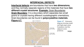
Understanding Interfacial Defects and Microscopy in Materials Science
Interfacial defects are boundaries separating regions with different crystal structures, like grain boundaries in polycrystalline materials. Microscopic examination distinguishes between macroscopic and microscopic dimensions, analyzed through optical and electron microscopy methods. Electron micros
0 views • 9 slides
Exploring Atomic Dimensions: Scanning Probe Microscopy
Delve into the world of nanoscale imaging with Scanning Probe Microscopy (SPM) techniques like Scanning Tunneling Microscopy (STM) and Atomic Force Microscope (AFM). Unlike optical microscopes, SPM methods break the diffraction limit by relying on intermolecular forces and quantum tunneling for unpa
0 views • 26 slides
Exploring Confocal Microscopy: Adding the Third Dimension
Delve into the world of confocal microscopy with Lecture 07 of Principles of Modern Microscopy. Learn about optical sectioning, wide-field imaging, confocal laser scanning, and more to enhance fluorescence in microscopy. Understand the methods of optical sectioning such as deconvolution, multi-photo
0 views • 65 slides
Fundamentals of Microscopy in Microbiology: An Insightful Journey
Explore the intriguing world of microscopy in microbiology, delving into the history, magnification, resolution, numerical aperture, and focal length. Uncover the evolution of microscopes and the fundamental concepts that underpin the science of microscopy. Gain a deeper understanding of how these t
0 views • 14 slides
Advancements in Medicine and Social Conditions in the Nineteenth Century
The nineteenth century marked a significant era in medical history with new cures, advancements, and improved social conditions. Progress in medical knowledge, understanding of disease causes, developments in surgery, and notable contributions from Pasteur, Koch, and Ehrlich transformed healthcare.
0 views • 33 slides
Transforming Geoscience Education with Remote Electron Microscopy
Expanding the use of online remote electron microscopy to enhance undergraduate geoscience education at Valencia College through a project focused on evaluating sands remotely using an operated scanning electron microscope. Students collect and analyze various sands using different tools and techniq
0 views • 10 slides
Understanding Transmission Electron Microscopy (TEM)
Transmission Electron Microscopy (TEM) is a powerful microscopy technique that uses a beam of electrons to create high-resolution images of ultra-thin specimens. It provides detailed information on topography, morphology, composition, and crystalline structure. Sample preparation for TEM is crucial,
0 views • 10 slides
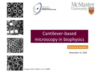
Understanding Cantilever-Based Microscopy in Biophysics
Cantilever-based microscopy in biophysics explores the operating principles of an Atomic Force Microscope (AFM). It delves into high-resolution imaging and nano-force testing, with applications in biophysics. AFM works by measuring the interaction between the atom on the tip and atoms in the sample,
0 views • 18 slides
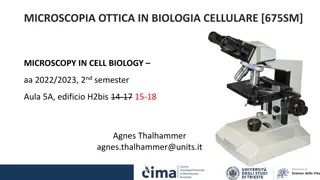
Microscopic Techniques in Cell Biology: Lessons and Labs Overview
Explore the fascinating world of optical microscopy in cellular biology, covering topics such as image formation, contrasting techniques, fluorescence microscopy, live-imaging techniques, and quantitative microscopy. Dive into the principles behind various microscopy techniques and experimental labs
0 views • 7 slides


