Advanced Microscopy Techniques and Applications in Cell Biology
Explore the diverse world of advanced microscopy techniques in cell biology, from confocal and super-resolution imaging to Fluorescence Resonance Energy Transfer (FRET) microscopy and Fluorescence Recovery After Photobleaching. Discover the latest methods, considerations, and applications for studying cellular structures and dynamics with cutting-edge imaging technologies.

Download Presentation

Please find below an Image/Link to download the presentation.
The content on the website is provided AS IS for your information and personal use only. It may not be sold, licensed, or shared on other websites without obtaining consent from the author.If you encounter any issues during the download, it is possible that the publisher has removed the file from their server.
You are allowed to download the files provided on this website for personal or commercial use, subject to the condition that they are used lawfully. All files are the property of their respective owners.
The content on the website is provided AS IS for your information and personal use only. It may not be sold, licensed, or shared on other websites without obtaining consent from the author.
E N D
Presentation Transcript
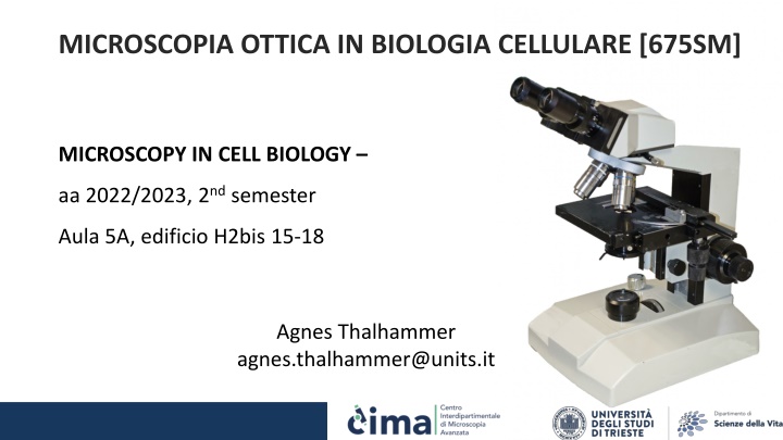
MICROSCOPIA OTTICA IN BIOLOGIA CELLULARE [675SM] MICROSCOPY IN CELL BIOLOGY aa 2022/2023, 2nd semester Aula 5A, edificio H2bis 15-18 Agnes Thalhammer agnes.thalhammer@units.it
CONCLUSIONS methods (TIRF, cofocal, SIM and 2 photon) use common dyes (good) confocal is easiest, most widely used best resolution obtainable only 100 nm (SIM) single molecule is problematic Consider sample thickness! 28 29
Advanced microscopy techniques CIMA SIM superresolution vs MP vs confocal cima.services@units.it confocal SIM MP Fixed sample yes yes yes Live sample not ideal yes dedicated incubation chamber with temperature, CO2and humidity regulation tens of mm preferably monolayer of cells yes to be set up customized to needs up to 2 mm tens of mm Sample thickness Wavelengths (nm) 405/488/561/640 405/488/561/640 tuneable, range 700-1000 nm strength Multichannel imaging for colocalisation Structural resolution and time series (1 fps) Penetration depth, time series (> 1 fps)
Fluorescence Resonance Energy Transfer (FRET) Microscopy 33
Fluorescence Resonance Energy Transfer (FRET) Microscopy 33
Fluorescence Resonance Energy Transfer (FRET) Microscopy 33
Fluorescence Resonance Energy Transfer (FRET) Microscopy 33
Fluorescence recovery after photobleaching Laser for photobleaching can be used on an epifluorescence or confocal microscope, usually with CCD camera, not PMT GFP dyes or photoconvertable dyes used 33
COMPARISON method excitation detection sectioning use Simple fluorescence samples High contrast images, optical sectioning Wide field Whole sample Whole sample No sectioning confocal Whole sample One z-plane 350-500nm Deep tissue imaging, optical sectioning 2-Photon One z-plane One z-plane 500-700nm FRET Protein interactions FRAP + photoactivation dynamics/mobility 405 laser (UV) Only bottom plane Only bottom plane TIRF Membrane dynamics 150-200nm
STIMULATED EMISSION DEPLETION (STED) MICROSCOPY (a) The process of stimulated emission. A ground state (S0) fluorophore can absorb a photon from the excitation light and jump to the excited state (S1). Spontaneous fluorescence emission brings the fluorophore back to the ground state. Stimulated emission happens when the excited-state fluorophore encounters another photon with a wavelength comparable to the energy difference between the ground and excited state. (b) The excitation laser and STED laser are combined and focused into the sample through the objective. A phase mask is placed in the light path of the STED laser to create a specific pattern at the objective focal point. (c) In the xy mode, a donut-shaped STED laser is applied with the zero point overlapped with the maximum of the excitation laser focus. With saturated depletion, fluorescence from regions near the zero point is suppressed, leading to a decreased size of the effective point spread function (PSF). 33
SUPER-RESOLUTION MICROSCOPY OF BIOLOGICAL SAMPLES. Schermelleh, Lothar, Rainer Heintzmann, and Heinrich Leonhardt. 2010. Aguide to super-resolution fluorescence microscopy. The Journal of Cell Biology 190 (2) (July 26): 165 - 175. doi:10.1083/jcb.201002021. 33
SINGLE MOLECULE LOCALISATION MICROSCOPY (SMLM) Induction of fluorescence emission of only a subset of molecules at a given time in order to localize each of them individually. By repeating this process, you can accumulate enough localizations to reconstruct a final super-resolved image. STORM and PALM are based on SMLM principle. The only difference is the way to induce the stochastic emission of the fluorophores: In STORM, fluorescent organic dyes (cyanines, rhodamines, etc.) together with a specific imaging buffer (abbelight s buffer) are used to allow blinking of the fluorescent molecules; In PALM, photoactivatable, photoconvertible or photoswitchable proteins are used (ex: PA-GFP, PA-mCherry, mEOS, mMAPLE, etc.); 33
PALM-Photoactivated localization microscopy invented by E Betzig and H Hess (2014 Nobel Prize for Chemistry) uses photoactivatable fluorophores to resolve spatial details of tightly packed molecules The laser stochastically activates fluorophores until all have emitted. Fluorophores emit for a short period but eventually bleach -> a more accurate view of positions can be obtained. 33
PALM-Photoactivated localization microscopy Signals from each fluorophore are still subject to the 300 nm diffraction of light limit. However, because each has been activated separately, the centre of mass can be calculated accurately The point spread function (PSF) is used to determine the location down to a resolution of 20 nm By mapping each of the more accurately defined points together, a complete super-resolution microscopy image can be compiled. 33
PALM-Photoactivated localization microscopy Three commonly used types of fluorophores are: Photoactivatable Fluorophores, e.g.: PAmCherry, PA-GFP, which emit light upon activation with UV. Photoconvertible Fluorophores, which change their emission spectrum upon activation with UV light (e.g.: mEOS proteins). Photoswitchable Fluorophores: Typically chemical dyes (e.g. Alexa Fluor 647, DyLight555) which can switch between dark, non- fluorescent and bright, fluorescent states repeatedly. 33
STORM-Stochastic Optical Reconstruction Microscopy https://downloads.microscope.healthcare.nikon.com/phase4/literature/Brochures/Super-Resolution_2CE-SCJK-3.pdf 33
STORM-Stochastic Optical Reconstruction Microscopy reconstructs a super- resolution image by combining the high- accuracy localization information of individual fluorophores https://downloads.microscope.healthcare.nikon.com/phase4/literature/Brochures/Super-Resolution_2CE-SCJK-3.pdf 33
STORM-Stochastic Optical Reconstruction Microscopy https://www.microscopyu.com/tutorials/sto chastic-optical-reconstruction-microscopy- storm-imaging 33
STORM-Stochastic Optical Reconstruction Microscopy 50 nm resolution https://downloads.microscope.healthcare.nikon.com/phase4/literature/Brochures/Super-Resolution_2CE-SCJK-3.pdf 33
SUPER-RESOLUTION MICROSCOPY OF BIOLOGICAL SAMPLES Schermelleh, Lothar, Rainer Heintzmann, and Heinrich Leonhardt. 2010. Aguide to super-resolution fluorescence microscopy. The Journal of Cell Biology 190 (2) (July 26): 165 - 175. doi:10.1083/jcb.201002021. 33


")
")
")
")


 MICROSCOPY")

")