Introduction to Microscopy: Care, Terms, and Operation
Explore the world of microscopy with proper care tips, essential terms like magnification and resolution, and detailed operation steps for using a microscope effectively. Learn about the components of a microscope and the process of preparing and viewing specimens at different magnification levels. Understand the significance of micrometers and nanometers in microscopy.
Download Presentation

Please find below an Image/Link to download the presentation.
The content on the website is provided AS IS for your information and personal use only. It may not be sold, licensed, or shared on other websites without obtaining consent from the author.If you encounter any issues during the download, it is possible that the publisher has removed the file from their server.
You are allowed to download the files provided on this website for personal or commercial use, subject to the condition that they are used lawfully. All files are the property of their respective owners.
The content on the website is provided AS IS for your information and personal use only. It may not be sold, licensed, or shared on other websites without obtaining consent from the author.
E N D
Presentation Transcript
Unit 1 Introduction to Microscopy
Microscopes Proper Care 1.)Expensive Carry with 2 hands 2.)Keep Glass parts clean! 3.)Never use Coarse adjustment with high Power or Oil Lens 4.)Start and stop with Low Power or Scanning Power
Terms 1.)Magnification ability of Microscope to enlarge image of object up to 1000X 3
2.)Resolution ability of Microscope to show detail clearly. 3.)Field of View(FOV) Diameter distance across light spot 4
4.)Micrometer 1000 micrometers in 1mm 5.)Nanometer -1000 nanometers in 1 micrometer Range of our Microscope =1 micrometer to 1000 micrometer. 5
Path of Light Bulb Diaphragm(adjusts light) Stage Hole Slide Specimen Cover Slip Objective Lens(Magnifies) Mirrors Eyepiece(Magnifies) Eyeball (Cornea to Lens to Retina to Optic Nerve to Brain) 6
Operation of the Microscope 1. Prepare slide 2. Put slide on stage 3. Turn light on 4. Use low power or scan power 1st 5. Use coarse adjustment 6. Use fine adjustment 7. Adjust diaphragm
8. Switch to High Power 9. *Fine Adjust Only 10. Adjust Diaphragm 11. Add Oil Drop 12. Turn to oil power 13. Fine Adjust 14. Adjust Diaphragm
Total Magnification (TM) 1. TM = eyepiece mag. X objective lens 2. Scanning Power TM = 10 x 4 = 40X 3. Low Power TM = 10 x 10 = 100X 4. High Power TM = 10 x 40 = 400X 5. Oil Power TM = 10 x 100 = 1000X
Types of Microscopes A. Light Microscope 1. Simple microscope one lens *Examples Magnifying glass + Leeuwenhoeks 2. Compound microscope two lenses: Ex. = ours
*More stuff about the compound microscope Real Image Created by objective lens Virtual image Image bouncing off mirror through eyepiece lens into the eyeball.
High Resolution occurs with shorter (blue) light waves. Since air refracts light, oil between slide and objective increases resolution. .2 Micrometer is the limit that light microscopes can resolve.
B. Stereoscopic Dissecting Microscope 1. Better for large and Opaque objects 2. Magnification ranges between 4X 40X 3. Has 2 Oculars (eyepieces) + 3d image 4. Can use reflected or transmitted light 5. real image
C. Bright Field Microscope *Most common Example = ours D. Dark Field Microscope - Bright specimen against a black field of view - poor internal details - Objective lens receives reflected light off of specimen
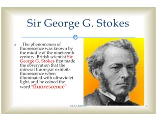
E.Phase Contrast Microscope shows great Internal Detail F. Interference Microscope shows great Internal Detail G. Fluorescent Microscope
H. Electron Microscope Page 67 1. Create the greatest magnified images with the greatest resolution 2. Very expensive $$$ 3. Cannot view live specimens 4. No colored images 5. Specimens pretreated with dyes 6. No direct observations viewed on a screen or photographed
Transmission Electron Microscope (TEM): a) Magnifies up to 1,000,000x b) Specimen Preparation = thin slice (20-100 nm) = staining c) Shows incredible internal detail
8. Scanning Electron Microscope (SEM)Produces super 3d image with great external detail a) Magnifies up to 100,000x
