Microscopic Techniques in Cell Biology: Lessons and Labs Overview
Explore the fascinating world of optical microscopy in cellular biology, covering topics such as image formation, contrasting techniques, fluorescence microscopy, live-imaging techniques, and quantitative microscopy. Dive into the principles behind various microscopy techniques and experimental labs to enhance your understanding of cell biology. Discover how microscopes work, different contrasting techniques, fluorescence principles, live-imaging methods, and image processing using ImageJ software. Get ready to immerse yourself in the intricate details of cellular structures and processes with hands-on learning experiences.
Download Presentation

Please find below an Image/Link to download the presentation.
The content on the website is provided AS IS for your information and personal use only. It may not be sold, licensed, or shared on other websites without obtaining consent from the author. Download presentation by click this link. If you encounter any issues during the download, it is possible that the publisher has removed the file from their server.
E N D
Presentation Transcript
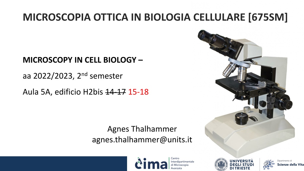
MICROSCOPIA OTTICA IN BIOLOGIA CELLULARE [675SM] MICROSCOPY IN CELL BIOLOGY aa 2022/2023, 2nd semester Aula 5A, edificio H2bis 14-17 15-18 Agnes Thalhammer agnes.thalhammer@units.it
MICROSCOPIA OTTICA IN BIOLOGIA CELLULARE [675SM] date lesson/lab aula time 14-15 (GF and BT), 16-17 (NS) 01/03/23 intro aula5A, H2bis 08/03/23 lesson1 aula5A, H2bis 15-18 15/03/23 lab1 sala microscopia F2, C1 15-18 22/03/23 lesson2 aula5A, H2bis 15-18 29/03/23 lab2 sala microscopia F2, C1 15-18 05/04/23 lesson3 aula5A, H2bis 15-18 19/04/23 lesson4 aula5A, H2bis 15-18 tbn lesson5 aula5A, H2bis tbn tbn lab3 sala microscopia F2, C1 tbn tbn lab4 CIMA center tbn 12 h lab + 16 h lessons
1. Lesson 1 - How a microscope works 1.1. Image formation 1.2. Magnification vs resolution 1.3. Numerical aperture and working distance 1.4. Objectives 1.5. Point-spread function and Airy disk 1.6. Optical abberations 2. Lab1 Contrasting techniques 2.1. Brightfield 2.2. Darkfield 2.3. Phase Contrast 2.4. Polarization Contrast 2.5. Differential Interference Contrast (DIC)
3. Lesson 2 - Flourescence microscopy 3.1. Flourescence principle 3.2. Absorption and Emission spectra - Stoke s shift 3.3. The flourescence microscope light sources, filter, dicroic mirror 3.4. Fluorophores 3.5. Staining with flourophores 3.6. Problems with flourescence imaging 3.7. Multichannel imaging 4. Lab2 - Flourescence microscopy
5. Lesson 3 Confocal, super-resolution and 2-photon microscopy 5.1. TIRF microscopy 5.2. Confocal microscopy 5.3. 2-photon microscopy 5.4. Superresolution microscopy 5.4.1. SIM microscopy 5.4.2. STED microscopy 5.4.3. PALM microscopy 5.4.4. STORM microscopy 5.5. FRET microscopy 5.6. FRAP microscopy lab4
6. Lesson4- Live-imaging techniques 6.1. Incubation 6.2. The life-imaging microscope 6.3. Contrasting techniques 6.4. Flourescent labelling of live cells 6.5. Resolution Speed Sensitivity 6.6. Examples
7. Lesson5 - Quantitative microscopy Imaging processing and analysis 7.1. Digital images 7.2. Resolution 7.3. Signal-to-noise 7.4. Sampling 7.5. Quantization 8. Lab3 Image processing and analysis using ImageJ 8.1. Histogram and LUTs 8.2. Noise, Filters and Background 8.4. ROIs and measurements 8.5. Threshold, watershed and particle analysis 8.6. Live imaging analysis
