Understanding the Compound Light Microscope and Its Applications
Explore the fundamental concepts of microscopy through Professor Diane Hilker's detailed lectures on the use and care of the compound light microscope. Dive into topics like magnification, resolution, and the practical application of microscopes in scientific research. Enhance your understanding of microscopy techniques and their importance in the field of microbiology.
Download Presentation

Please find below an Image/Link to download the presentation.
The content on the website is provided AS IS for your information and personal use only. It may not be sold, licensed, or shared on other websites without obtaining consent from the author. Download presentation by click this link. If you encounter any issues during the download, it is possible that the publisher has removed the file from their server.
E N D
Presentation Transcript
Exp. 1: Introduction to the Microscope I. Exp. 2: Survey of Microbes II. III. Exp. 3: Collection of Microbes
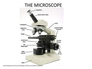
Purpose: the use & care of the compound light microscope Purpose: To review Fig. 3.1 Textbook
Compound Binocular Light Microscope (2) Sources of Magnification: (2) Sources of Magnification: Eyepiece or Ocular (10x) Objectives (4): Scanning Power: 4 x Low Power: 10x High Power: 40x Oil Immersion: 100x Parfocal another with minimal focusing Compound Binocular Light Microscope Parfocal: : ability to go from one objective to
Total Magnification: TM TM=Magnification of Eyepiece X Magnification of Objective Total Magnification: TM Eyepiece Magnification 10X Eyepiece Magnification Objective Magnification 4X Objective Magnification TM TM Scanning 40X Low 10X 10X 100X High Dry 10X 40X 400X Oil Immersion 10X 100X 1000X
Resolution or Resolving Power (RP) Ability to distinguish detail clearly Resolution or Resolving Power (RP) To be able to tell 2 points as separate points and not one point RP = Wavelength of Light = ------- 2 X Numerical Aperture 2NA
Wavelength of light (nm) Red light = 700 nm Blue light = 400 nm Wavelength of light (nm) Red light = 700 nm Blue light = 400 nm RP 700 nm = 350 nm 2(1) = 400 nm 2(1) Lower the resolution, better the clarity Blue filter provides the best resolution with a halogen light bulb Blue filter NOT needed with microscopes that have a LED light bulb RP red red = = 700 nm 2(1) blue = = 350 nm RP RP blue 400 nm = 200 nm 2(1) Lower the resolution, better the clarity Blue filter provides the best resolution with a halogen light bulb Blue filter NOT needed with microscopes that have a LED light bulb = 200 nm
Numerical Aperture (NA): of light that enters the lens so as to see fine detail. Two things make up NA: Angular Aperture through the lenses & filters of the condenser & into the objective (Constant) Refractive Index: a medium Refractive Index of Air = 1.0 Refractive Index of Oil = 1.5 Numerical Aperture (NA): describes the cone Angular Aperture: angle of light as it goes Refractive Index: how light travels through
RP RP air air= = 400 nm 2(1.0) 400 nm = 200 nm 2(1.0) = 200 nm RP RPoil oil= = 400 nm 2(1.5) 400 nm = 133 nm 2(1.5) = 133 nm Better resolution with oil
Exp. 1: Introduction to the Microscope I. Exp. 2: Survey of Microbes II. III. Exp. 3: Collection of Microbes
Purpose: microscope & to view various microbes Purpose: To become familiar with using a Wet Mount Focus on edge of coverslip Scanning-dim light using diaphragm Move toward center of slide Observe under Low & High Powers Slides will dry out quickly Wet Mount: observing living cells
4 Slides: Largest to smallest microorganisms 4 Slides: Largest to smallest microorganisms 1. Pond Water: algae 1. Pond Water: algae- -much variation much variation
2. Protozoa: that move by different methods that belong to the Protista kingdom. 2. Protozoa: single celled eukaryotic microbes Pseudopods: Pseudopods: false feet Amoeba Cilia Paramecium Cilia Flagella Flagella Euglena
3. Yeast: that belong to the Fungi kingdom 3. Yeast: single celled eukaryotic microbes Ovoid & irregular Budding: method of reproduction Brownian movement Smaller than protozoa Larger than bacteria
4. Bacteria (Hay infusion ): prokaryotic microbes that belong to the Eubacteria Domain. Must view under 400x Very small Motile & non-motile Looks like specks of sand Hard to discern shape Smaller than yeast & protozoa Protozoa may be present in the sample 4. Bacteria (Hay infusion ): single celled
Exp. 1: Introduction to the Microscope I. Exp. 2: Survey of Microbes II. III. Exp. 3: Collection of Microbes
Purpose: the environment for observation Purpose: To collect and grow microbes from Procedure to be described by lab instructor
