Deep Learning for Plant Disease Resistance Analysis
Utilizing deep learning facilitated microscopy, a research team led by Hening Cui from Columbia University aims to dissect durable resistance to plant diseases. The project focuses on segmenting hyphal networks of fungal and host plant cells using a deep convolutional neural network architecture called DeepXScope. The goals include running the pipeline on various datasets, testing it on different microscopy platforms, and producing a publication-quality graphical schema representing the pipeline and user interaction.

Uploaded on Sep 26, 2024 | 6 Views
Download Presentation

Please find below an Image/Link to download the presentation.
The content on the website is provided AS IS for your information and personal use only. It may not be sold, licensed, or shared on other websites without obtaining consent from the author.If you encounter any issues during the download, it is possible that the publisher has removed the file from their server.
You are allowed to download the files provided on this website for personal or commercial use, subject to the condition that they are used lawfully. All files are the property of their respective owners.
The content on the website is provided AS IS for your information and personal use only. It may not be sold, licensed, or shared on other websites without obtaining consent from the author.
E N D
Presentation Transcript
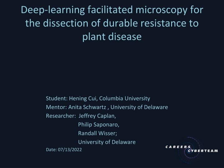
Deep-learning facilitated microscopy for the dissection of durable resistance to plant disease Student: Hening Cui, Columbia University Mentor: Anita Schwartz , University of Delaware Researcher: Jeffrey Caplan, Philip Saponaro, Randall Wisser; University of Delaware Date: 07/13/2022
Background High-speed confocal microscopy on plant-fungal interactions Segmentation manually, infeasible for large amounts of data developing separate algorithms to extract individual features Maize leaf with C. heterostrophus (green) S: stoma E: lower epidermis V: Vascular bundle M: mesophyll Minker, K. R ., et al., 2018
Method DeepXScope single deep convolutional neural network architecture automatically segmenting hyphal networks of the fungal and host plant cell Github: https://github.com/drmaize/compvision Saponaro, P. et al.,2017
Process schema Saponaro, P. et al.,2017
Result Connect gaps Raw 3D image Quantification Skeleton connection Surface Estimation Reduce noise 2D image with depth Extract pixel with interest Threshold Skeletonizing Segmentation Minker, K. R ., et al., 2018 ; Saponaro, P. et al.,2017
Goals Familiar with DeepXScope pipeline functions, installation, and how to process images, writing updates to the public README as needed Run DeepXScope on existing and incoming datasets to generate standardized output to be shared with the research team Test the pipeline on data collected from two other microscopy platforms Produce a publication quality graphical schema describing the pipeline and user interaction
Timeframe Start date: June 14th End date: November 6th
What I hope to learn Deep learning pipeline functioning Server command line CNN machine learning model
Goals for Next Month Generate both manually annotated and simulated ground-truth data on one dataset Assess accuracy of the pipeline s segmentation routines using precision and recall metrics
Help Provided Accounts sponsored by UD's ECE/CIS to access UD's Community Cluster Caviness to install DeepXScope for remote access vs using a local laptop/computer install All the research team s kindly help
References Minker, K. R., Biedrzycki, M. L., Kolagunda, A., Rhein, S., Perina, F. J., Jacobs, S. S., ... & Caplan, J. L. (2018). Semiautomated confocal imaging of fungal pathogenesis on plants: microscopic analysis of macroscopic specimens. Microscopy research and technique, 81(2), 141-152. Saponaro, P., Treible, W., Kolagunda, A., Chaya, T., Caplan, J., Kambhamettu, C., & Wisser, R. (2017). Deepxscope: Segmenting microscopy images with a deep neural network. In Proceedings of the IEEE conference on computer vision and pattern recognition workshops (pp. 91-98).