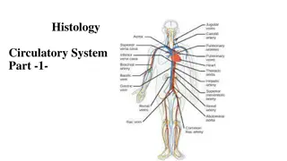
Understanding the Heart: Structure, Function, and Circulation

Explore the intricate details of the heart, from its anatomy and functions to interesting facts and the flow of blood. Discover how this vital organ pumps oxygenated blood to the body and facilitates gas exchange, while learning about its four chambers, valves, and the critical role it plays in cardiovascular health.

Download Presentation

Please find below an Image/Link to download the presentation.
The content on the website is provided AS IS for your information and personal use only. It may not be sold, licensed, or shared on other websites without obtaining consent from the author. Download presentation by click this link. If you encounter any issues during the download, it is possible that the publisher has removed the file from their server.
E N D
Presentation Transcript
THE HEART OF THE MATTER Structure and Function of the Heart
5 INTERESTING FACTS ABOUT THE HEART Each day, your heart beats 100,000 times. Each minute, your heart pumps 1.5 gallons of blood. Heart disease is the #1 cause of death. A normal heart valve is about the size of a half dollar coin. The largest heart ever recorded belonged to a grey whale. http://www.culinaryschools.org/clipart/drinks/milk-gallon.gif
FUNCTIONS OF THE HEART Pumps oxygenated and nutrient-rich blood to the body through blood vessels Pumps deoxygenated blood, containing wastes, to the lungs, where gas exchange to the outside environment occurs
ANATOMY OF THE HEART Located under rib cage and in between the lungs Size varies depending on age, size, and condition of the heart On average, the heart is about the size of that person s clenched fist
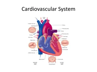
FOUR CHAMBERS OF THE HEART Right Atrium Right Ventricle Left Atrium Left Ventricle http://planetearth.nerc.ac.uk/images/uploaded/custom/heart_interior-c.gif
ATRIA SEPTUM The upper two chambers of the heart Receive and collect blood Muscle that divides the heart into right and left halves http://planetearth.nerc.ac.uk/images/uploaded/custom/heart_interior-c.gif VENTRICLES The lower two chambers of the heart Pump blood out of the heart
VALVES http://planetearth.nerc.ac.uk/images/uploaded/custom/heart_interior-c.gif 4 Heart Valves Tricuspid Valve Pulmonary Semilunar Valve Mitral (Biscuspid) Valve Aortic (Semilunar) Valve Purpose: prevent backflow of blood, keep blood flowing in one direction
FLOWOF BLOOD 1. Deoxygenated blood from the upper and lower body flows through the superior and inferior vena cava. http://planetearth.nerc.ac.uk/images/uploaded/custom/heart_interior-c.gif
FLOWOF BLOOD 2. The superior and inferior vena cava empty blood into the right atrium. http://planetearth.nerc.ac.uk/images/uploaded/custom/heart_interior-c.gif
FLOWOF BLOOD 3. Blood from the right atrium passes through the tricuspid valve. http://planetearth.nerc.ac.uk/images/uploaded/custom/heart_interior-c.gif
FLOWOF BLOOD 4. Blood passes through the tricuspid valve into the right ventricle. http://planetearth.nerc.ac.uk/images/uploaded/custom/heart_interior-c.gif
FLOWOF BLOOD 5. Blood from the right ventricle passes through the pulmonary semilunar valve. http://planetearth.nerc.ac.uk/images/uploaded/custom/heart_interior-c.gif
FLOWOF BLOOD 6. Blood flows through the pulmonary semilunar valve into the right and left pulmonary arteries. http://planetearth.nerc.ac.uk/images/uploaded/custom/heart_interior-c.gif
FLOWOF BLOOD http://www.mhhe.com/biosci/esp/2001_gbio/folder_structure/an/m7/s3/assets/images/anm7s3_1.jpg 7. Pulmonary arteries take blood to the lungs for gas exchange. In the lung capillaries, blood picks up oxygen and transfers carbon dioxide to the lungs for exhalation. Blood becomes oxygenated.
FLOWOF BLOOD 8. Right and left pulmonary veins bring the blood back to the heart. http://planetearth.nerc.ac.uk/images/uploaded/custom/heart_interior-c.gif
FLOWOF BLOOD 9. Pulmonary veins empty blood into the left atrium. http://planetearth.nerc.ac.uk/images/uploaded/custom/heart_interior-c.gif
FLOWOF BLOOD 10. The blood from the left atrium flows through the mitral (or bicuspid) valve. http://planetearth.nerc.ac.uk/images/uploaded/custom/heart_interior-c.gif
FLOWOF BLOOD 11. After passing through the mitral valve, blood enters the left ventricle. http://planetearth.nerc.ac.uk/images/uploaded/custom/heart_interior-c.gif
FLOWOF BLOOD 12. Blood from the left ventricle passes through the aortic semilunar valve. http://planetearth.nerc.ac.uk/images/uploaded/custom/heart_interior-c.gif
FLOWOF BLOOD 13. After passing through the aortic semilunar valve, the blood enters the aorta and is then pumped to the rest of the body. http://planetearth.nerc.ac.uk/images/uploaded/custom/heart_interior-c.gif
FLOWOF BLOODIN ACTION YouTube Video - How a Normal Heart Pumps Blood
http://www.mhhe.com/biosci/esp/2001_gbio/folder_structure/an/m7/s3/assets/images/anm7s3_1.jpghttp://www.mhhe.com/biosci/esp/2001_gbio/folder_structure/an/m7/s3/assets/images/anm7s3_1.jpg CIRCULATION Pulmonary circuit: movement of blood from the heart to the lungs and back to the heart
http://www.mhhe.com/biosci/esp/2001_gbio/folder_structure/an/m7/s3/assets/images/anm7s3_1.jpghttp://www.mhhe.com/biosci/esp/2001_gbio/folder_structure/an/m7/s3/assets/images/anm7s3_1.jpg CIRCULATION Systemic circulation: movement of blood from the body to the heart and back to the body Why do you think the left side of the heart is larger than the right? Answer: Because the left side has to pump blood further!
CONTRACTION Systole - contract Atrial Systole: when the atria contract and pump blood into the ventricles Ventricular Systole: when the ventricles contract and pump blood out of the heart to the lungs or body Diastole - relax When the atria and ventricles relax and start to fill with blood
DID YOU KNOW? Did you know that even outside of the body, the heart will continue to beat? Why do you think this is? This characteristic is called myogenic control. Each heart beat is caused by an electrical signal from within heart muscle itself.
ELECTRICAL SYSTEM AKA the Cardiac Conduction System Consists of three parts: 1. Sinoatrial (SA) node 2. Atrioventricular (AV) node 3. Bundle of His and Purkinje fibers
ELECTRICAL SYSTEM 1. Electrical signal starts at the SA node as blood fills the right atrium. This signal causes the atrium to contract. The SA node sets the pace of the heart, so it is also called the pacemaker. 2. Signal arrives at the AV node as blood fills the ventricles. 3. Signal moves along the Bundle of His and along the walls of the ventricles. The Bundle of His divides into right and left branches and then to Purkinje fibers. The ventricles contract. 4. Signal passes and ventricles relax.
ELECTRICAL SYSTEMIN ACTION YouTube Video - Electrical Conduction in Heart
EXAMPLESOF HEART DISEASES/CONDITIONS Congestive Heart Failure The heart is too weak or stiff to pump blood effectively. Myocardial Infarction (Heart attack!) The coronary artery is blocked so blood cannot supply the heart with oxygen, and heart muscle dies. Atrial Fibrillation Abnormal electrical impulses in the atrium cause irregular heart beat.
WAYSTO PREVENT HEART DISEASE Don t smoke or use tobacco Exercise 30 minutes a day Eat a heart healthy diet Fruits Vegetables Whole grains Nuts Fish No saturated or trans fats
REFERENCES http://www.nhlbi.nih.gov/health/health- topics/topics/hhw/ http://www.webmd.com/heart/picture-of-the-heart http://www.mayoclinic.org/diseases- conditions/heart-disease/in-depth/heart-disease- prevention/art-20046502 http://health.clevelandclinic.org/2013/07/19- amazing-facts-about-your-heart-infographic/
3-2-1 On a piece of notebook paper, write 3 things you learned about the heart 2 things you have questions about 1 thing you wish for me to know