Overview of Veins and Venous Circulation in the Body

Veins play a crucial role in the circulatory system by carrying deoxygenated blood back to the heart. This lecture covers the general principles of veins, the anatomy of major veins like the superior and inferior vena cavae, tributaries, and their roles in different parts of the body. It also discusses the hepatic portal vein, venous drainage from the head and neck, and the importance of anastomoses in venous circulation.

Download Presentation

Please find below an Image/Link to download the presentation.
The content on the website is provided AS IS for your information and personal use only. It may not be sold, licensed, or shared on other websites without obtaining consent from the author. Download presentation by click this link. If you encounter any issues during the download, it is possible that the publisher has removed the file from their server.
E N D
Presentation Transcript
Objectives At the end of the lecture, the student should be able to: Define the vein and understand the general principle of the veins. Describe the role of the superior and inferior vena cavae & list the tributaries of each vein. List major veins in the head & neck, thorax, abdomen and upper & lower extremities. Describe the hepatic portal vein and list its tributaries.
General principles about Veins Veins are the vessels that carry blood towards the heart. All veins, carry deoxygenated blood, except the pulmonary and umbilical arteries, which carry oxygenated blood from the lungs (postnatal) and from the placenta (prenatal) respectively Veins have valves that allow unidirectional blood flow. The flow of blood depends on the peripheral muscular activity Veins begin as venules, which unite into vessels of increasing size to form veins Arranged as superficial and deep veins, draining the superficial and deep parts of the body respectively. Deep veins are paired with arteries. Superficial veins usually have names unrelated to those of arteries, because there are very few superficial arteries. Vary considerably in their locations and branching Communicate with each other forming extensive anastomoses which provides for collateral return blood flow in case of venous obstruction.
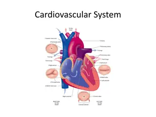
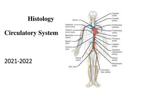
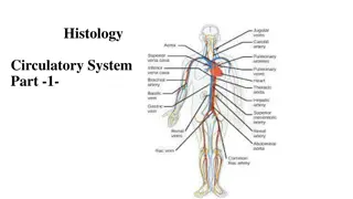
Venous blood from the body is drained into the right atrium by two major veins: The superior vena cava, which brings blood from all parts above the diaphragm: the head, neck, brain, upper limbs, and much of the chest and lungs. The inferior vena cava, whichbrings blood from all parts below the diaphragm: abdomen, pelvis, perineum and lower limbs.
The Superior Vena Cava Formed by the union of the 2 (right & left) brachiocephalic veins. Each brachiocephalic vein is formed by the union of internal jugular and subclavian veins. Drains into the right atrium. Tributaries Azygos vein. Pericardial veins. Mediastinal veins.
The Inferior Vena Cava Formed by the union of the two common iliac veins, at the level of L5. Tributaries Inferior phrenic Right supra renal Renal Hepatic veins Lumbar veins Right gonadal (left gonadal drains into left renal vein)
Veins of the Head and Neck Superficial veins Deep veins The superficial veins of the head & neck drain into the paired external jugular veins. The external jugular veins empty into the subclavian veins.
The deep veins of the head & neck draining the: Brain, lie in the cranial cavity. These are dural sinuses large, elongated chambers formed between layers of the dura mater Deep structures in the neck ultimately drain into the internal jugular veins. Small emissary veins connect the superficial veins with the dural venous sinuses, a fact of clinical interest as a possible avenue for infections to enter the cranial cavity.
Veins of the Upper Limb Superficial: Cephalic Basilic Median cubital Deep: Venae comitantes of the Radial,ulnar and brachial arteries. Axillary vein Subclavian vein: continuation of axillary, at the outer border of the first rib
Veins of the Lower Limb Superficial: Great saphenous Small saphenous Deep: Venae comitantes of the anterior tibial artery. Venae comitantes of the posterior tibial artery. Popliteal Femoral External Iliac
Veins of the Thorax The bronchial, esophageal and pericardial veins drain into superior vena cava or azygos vein The posterior intercostal veins drain into azygos and hemiazygos veins The anterior intercostal veins drain into the internal thoracic veins which drains into the corresponding brachiocephalic veins
Veins of the Abdomen Veins from the abdominal walls drain into the inferior vena cava Veins returning from the digestive tract, pancreas, and spleen merge to form the hepatic portal vein, which does not join the inferior vena cava. Instead, it redivides and flows into a network of sinusoids in the liver. This collection of two capillary beds (the intestinal capillaries and the hepatic sinusoids) and the hepatic portal vein connecting them is called the hepatic portal circulation
Hepatic Portal Vein Drains the GIT, spleen, pancreas and gall bladder Delivers this blood to the liver Formation: Formed by the union of the superior mesenteric vein and the splenic vein. The Inferior mesenteric vein drains into the splenic vein Liver is drained by hepatic veins which open into the inferior vena cava
Thank you Thank you