Understanding the Circulatory System: A Detailed Overview

The circulatory system plays a vital role in transporting nutrients, oxygen, and other essential substances throughout the body. It consists of the blood vascular system (arteries, arterioles, veins, capillaries, and the heart) and the lymphatic vascular system. Understanding the general structure of blood vessels, with their three layers (intima, media, adventitia), is key to comprehending how circulation works. Explore the intricacies of this system to appreciate its importance in maintaining homeostasis and overall health.

Download Presentation

Please find below an Image/Link to download the presentation.
The content on the website is provided AS IS for your information and personal use only. It may not be sold, licensed, or shared on other websites without obtaining consent from the author. Download presentation by click this link. If you encounter any issues during the download, it is possible that the publisher has removed the file from their server.
E N D
Presentation Transcript
Histology Circulatory System 2021 2021- -2022 2022
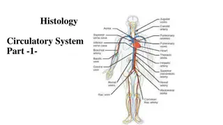
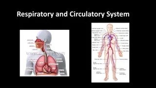
Circulatory System Circulatory System The circulatory system is an organ system that permits blood and lymph circulation to transport nutrients (such as amino acids and electrolytes), oxygen, carbon dioxide, hormones, blood cells, etc. to and from cells in the body to nourish it and help to fight diseases, stabilize body temperature and pH, and to maintain homeostasis. The vascular system is a continuous network of tubes or vessels that distributes blood throughout the body and returns it the heart.
The circulatory system consist of two systems: The circulatory system consist of two systems:- - 1-Blood vascular system or cardiovascular system. 2-Lymphatic vascular system. A. The cardiovascular system A. The cardiovascular system The blood vascular system is composed of the following: 1 1.Arteries .Arteries 2 2. Arterioles . Arterioles 3 3.Veins 4 4.Capillaries .Capillaries 5 5.Heart .Heart 6 6.Venules .Veins .Venules
B. The lymph vascular system B. The lymph vascular system 1. Blind ended lymphatic capillaries Blind ended lymphatic capillaries that collect lymph fluid from tissues. 2. larger lymphatic vessels larger lymphatic vessels that connect with one and other and finally empty collected lymph into large veins in the neck where the lymphatic and cardiovascular systems merge.
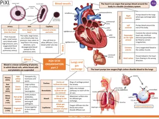
General Structure of Blood Vessels General Structure of Blood Vessels Blood vessels of various sizes and should be aware that the histological appearances of vessels of different sizes (arterioles, arteries) and different types (arteries ,veins) are different from each other. These differences are the result of quantitative variations of a common structural pattern that can be seen in all blood vessels with the exception of capillaries, i.e...
the division of the walls of the blood vessels into three layers or tunics . These are arranged into three concentric layers: intima, media and adventitia. The intima is the inner layer abutting the vessel lumen. The adventitia is the outer layer abutting the perivascular soft tissue. The media is sandwiched between the intima and adventitia
1 - -The The intima intima is the thinnest layer. It is composed of a single layer of endothelial cells and a small amount of sub endothelial connective tissue. The intima is separated from the media by a dense elastic membrane called the internal elastic lamina. 2 2- -The The media media is the thickest layer and provides structural support, vasoreactivity and elasticity. It is composed of smooth muscle cells, elastic fibers and connective tissue, which vary in amount depending on the type of vessel. Smooth muscle cells contract (vasoconstriction) or relax (vasodilatation), which is controlled by autonomic nerves (nervivasorum) and local metabolic factors
3 3- -The The adventitia adventitia is composed of connective tissue, nutrient vessels (vasa vasorum) and autonomic nerves (nervivasorum). The intima and inner part of the media are nourished by diffusion of oxygen and nutrients from blood in the lumen, and the adventitia and outer part of the media are nourished by vasa vasorum.
Arteries Arteries The walls of arteries are thicker than that of veins to withstand pulsatile flow and higher blood pressures. As arteries become smaller, wall thickness gradually decreases but the ratio of wall thickness to lumen diameter increases . Arteries are divided into three types according to size and function. The constituents of the media of these vessels differ in their relative amounts accordingly.
1 1- -Large elastic arteries Large elastic arteries (aorta, large aortic branchesand pulmonary arteries) the media is abundant in elastic fibers that allow it to expand with systole and recoil during diastole, thereby propelling blood forward. 2 2- -Medium Medium- -sized muscular arteries sized muscular arteries (other aortic branches, coronary and renal arteries) the media is abundant in smooth muscle cells that vasoconstriction or vasodilation, thereby controlling lumen diameter and regional blood flow. 3 3- -Small arteries and arterioles Small arteries and arterioles (in the substance of organs and tissues) the media is abundant in smooth muscle cells that vasoconstriction or vasodilation. In vessels of this size, smooth muscle contraction causes dramatic changes in lumen diameter, thereby controlling systemic blood pressure as well as regional blood flow.
Veins Veins The walls of veins are thinner than the walls of arteries, while their diameter is larger. In contrast to arteries, the layering in the wall of veins is not very distinct. The tunica intima is very thin. Only the largest veins contain an appreciable amount of sub endothelial connective tissue. Internal and external elastic laminae are absent or very thin. The tunica media appears thinner than the tunica adventitia, and the two layers tend to blend into each other.
The appearance of the wall of veins also depends on their location. The walls of veins in the lower parts of the body are typically thicker than those of the upper parts of the body, and the walls of veins which are embedded in tissues that may provide some structural support are thinner than the walls of unsupported veins.
VenaCava VenaCava The inferior vena cava is the largest vein in the human body. It collects blood from veins serving the tissues inferior to the heart and returns this blood to the right atrium of the heart. There are two in humans, the inferior vena cava (carrying blood from the lower body) and the superior vena cava (carrying blood from the head, arms, and upper body). Although the vena cava is very large in diameter, its walls are incredibly thin due to the low pressure exerted by venous blood.
Comparison between a vein and artery Artery Vein Thick wall Thin wall 1. 1. shape less deformed shape flatted 2. 2. tunica intina crinkled Smooth 3. 3. three distinct layers layering indistinct 4. 4. tunica media prominent Weak 5. 5.
Capillaries Capillaries are small, normally around 3-4 m, but some capillaries can be 30-40 m in diameter. The largest capillaries are found in the liver. Capillaries Capillaries connect arterioles arterioles to venules the exchange of nutrients and wastes between the blood and the tissue cells, together with the interstitial fluid. This exchange occurs by passive diffusion and by pinocytosis which means 'cell drinking. venules. They allow
Types of capillaries Types of capillaries 1 1- -Fenestratedcappillaries Fenestratedcappillaries: : Have the same structure of non fenestrated but several circular opening in their endothelium cell are closed by diaphragm.This type capillary found in kidney and endocrine gland.
2 2- -Sinusoid or discontinuous capillary Sinusoid or discontinuous capillary A large capillaries with diameter of (30-40)pm or more. It consists of cells which are irregular in shape and discontinuous basal membrane. The endothelium is not connected by desmosome and associated with macrophagocytic or (Kupffer cells). This type is found in spleen, bone marrow, and liver.
3 3- -Non fenestrated Non fenestrated ( (Continuous capillaries) Continuous capillaries) Are most common in body consist the wall endothelial cell resting on basal membrane and contain pericyte and pinocytotic vessel. This type capillary found in skin and muscle. Are formed by "continuous" endothelial cells and basal lamina. The endothelial cell and the basal lamina do not form openings, which would allow substances to pass the capillary wall without passing through both the endothelial cell and the basal lamina. Both endothelial cells and the basal lamina can act as selective filters in continuous capillaries.
Heart Heart The heart is a muscular organ in humans and other animals, which pumps blood through the blood vessels of the circulatory system. Blood provides the body with oxygen and nutrients, and also assists in the removal of metabolic wastes. The heart is located in the middle compartment of the mediastinum in the chest, the heart is a pump with four chambers and valves that maintain a one-way flow of blood. The wall of heart consists of three layers that are homologous to the three tunic of blood vessels.
Tunica Intima: ( Endocardium) Tunica Intima: ( Endocardium) The Endocardium is composed of a continuous endothelial lining, with attendant basement membrane, that lies on top of a layer of collagen. The endothelium of the heart is continuous with that of the aorta, vena cava, pulmonary artery, and pulmonary veins, Purkinje Fibers ( impulse conducting fibers ,it is large modified of muscle cell).
Tunica Media: ( Myocardium) Tunica Media: ( Myocardium) The myocardium is a variably-sized layer of cardiomyocytes that lies between the endocardium and epicardium and makes up the bulk of the heart's mass. Cardiomyocytes are arranged end to end, in a branching network of fibers and are attached to one another by specialized sections of membrane known as intercalated discs. The membrane at intercalated discs possess a large number of gap junctions which allow nearly free passage of ions between cardiomyocytes which is critical for cardiac action potential propagation.
Tunica Adventitia: (Epicardium) Tunica Adventitia: (Epicardium) The Epicardium of the heart is lined by a layer of flat mesothelial cells that lie on top of a layer of collagenous tissue. The epicardium makes up the visceral pericardium and serves as a lubricated surface which allows for the free movement of the heart within the pericardium. The outer, parietal pericardium also possesses a layer of mesothelium but is not considered a histological component of the heart itself.
Heart also have four valves composed of connective tissue layered with endothelium on each side, valves consist of three layes: Spongiosa (loose collagen),fibrosa(dense of connective tissue), Ventricularis


")
")
")