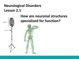
Overview of the Nervous System Components and Functions
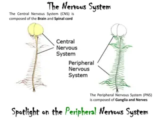
The nervous system is comprised of the central nervous system (CNS) and the peripheral nervous system (PNS). The CNS acts as the command center, interpreting sensory information and coordinating responses. The PNS conveys information to and from the CNS through somatic and visceral sensory neurons, as well as somatic and visceral motor neurons. The PNS further branches into the somatic and autonomic nervous systems, with divisions such as the sympathetic and parasympathetic systems. Within the CNS, structures like the cerebrum with its lobes play vital roles in information processing.

Download Presentation

Please find below an Image/Link to download the presentation.
The content on the website is provided AS IS for your information and personal use only. It may not be sold, licensed, or shared on other websites without obtaining consent from the author. Download presentation by click this link. If you encounter any issues during the download, it is possible that the publisher has removed the file from their server.
E N D
Presentation Transcript
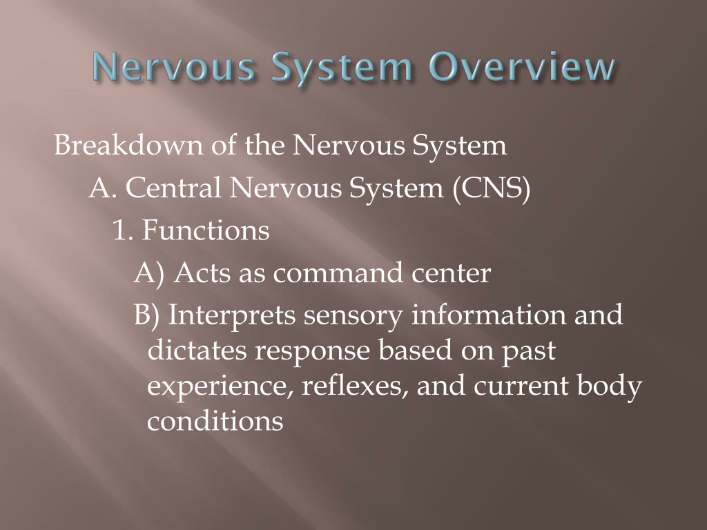
Breakdown of the Nervous System A. Central Nervous System (CNS) 1. Functions A) Acts as command center B) Interprets sensory information and dictates response based on past experience, reflexes, and current body conditions
B. Peripheral Nervous System (PNS) 1. Functions A) Conveys information to and from CNS 2. Components A) Somatic sensory (afferent) neurons 1) Carry impulses from receptors in skin, skeletal muscle, and joints to the CNS
B) Visceral sensory neurons 1) Carry impulses from receptors within the visceral organs to the CNS C) Somatic motor (efferent) neurons 1) Carry impulses from the CNS to skeletal muscles D) Visceral motor neurons 1) Carry impulses from the CNS to smooth muscle, cardiac muscle, and glands
C. Divisions of PNS 1. Somatic NS A) Consists of somatic sensory neurons and somatic motor neurons B) Voluntary nervous system 2. Autonomic NS A) Consists of visceral sensory neurons and visceral motor neurons
B) Involuntary nervous system C) 2 branches 1) Sympathetic fight-or-flight system a) Stimulates most effectors 2) Parasympathetic energy restoration/conservation system a) Inhibits most effectors
The Central Nervous System A. Structures of the CNS 1. Cerebrum A) Divided into 2 hemispheres 1) Each consists of gyri (elevated areas), sulci (shallow depressions) and fissures
B) 5 lobes 1) Frontal, parietal, occipital, temporal, and insula C) Important structures 1) Longitudinal fissure (right & left hemispheres) 2) Transverse fissure (cerebrum & cerebellum) 3) Central sulcus (frontal & parietal lobes)
a) Precentral gyrus (within frontal lobe) b) Postcentral gyrus (within parietal lobe) 4) Parieto-occipital sulcus (parietal & occiptal lobes) 5) Lateral sulcus (temporal & frontal/parietal lobes)
D) Cerebral cortex "conscious mind" 1) Composed of gray matter 2) Involved with memory, reasoning, intelligence, etc... 3) Contrilateral
4) Exhibits hemisphere dominance a) Left hemisphere most functions; 90% of population b) Right hemisphere artistic & musical qualities; left-handed
5) 3 main functional areas a) Motor areas i) Primary motor cortex (a) Found in precentral gyrus (b) Responsible for conscious movement of skeletal muscles
ii) Premotor cortex (a) Lies anterior to primary motor cortex (b) Responsible for learned motor skills that are repeated or patterned (ex. typing) iii) Broca s area (a) Lies anterior & inferior to premotor cortex (b) Involved in speech production (c) Only in one hemisphere (usually left)
iv) Frontal eye field (a) Lies anterior to premotor cortex and superior to Broca s area (b) Responsible for voluntary eye movements
b) Sensory areas i) Primary somatosensory cortex (a) Lies in postcentral gyrus (b) Allows for spatial discrimination ii) Somatosensory association cortex (a) Lies posterior to primary somatosensory cortex
(b) Integrates and analyzes somatic sensory inputs (i.e. pain, touch, temp, etc.) to produce an understanding of what is being felt iii) Visual area (a) Located within occipital lobes iv) Auditory area (a) Found in temporal lobes
v) Olfactory area (a) Found in temporal lobes in regions known as the uncus vi) Gustatory area (a) Found in parietal lobe
c) Association areas i) Prefrontal cortex (a) Found in anterior portions of frontal lobe (b) Involved with intellect, complex learning, and personality
ii) Gnostic area (a) Found in undefined areas of parietal, temporal, and occipital lobes (b) Only one per hemisphere (c) Receives input from all sensory association areas (d) Sends input to prefrontal cortex which adds emotions
iii) Language areas (a) Surround lateral sulcus in left hemisphere (b) 4 defined areas (i) Wernick s area (a) Associated with sounding out unfamaliar words
(ii) Brocas area (a) Associated with speech production (iii) Lateral prefrontal cortex (a) Associated with language comprehension and word analysis (iv) Lateral & ventral temporal lobes (a) Coordinates auditory & visual aspects of language
E) Cerebral white matter 1) Lies deep to cortex 2) Responsible for communication between cortical areas and also between the cortex and lower CNS centers 3) 3 types a) Commissures connect the right & left sides of the cerebrum
b) Association fibers transmit within the same hemisphere c) Projection fibers run to and from lower brain areas F) Basal nuclei 1) Bundles of subcortical gray matter deep within white matter 2) Control large automatic skeletal muscle contractions and produce dopamine
2. Diencephalon central core of brain; covered by cerebrum; 3 paired structures A) Thalamus connected by massa intermedia 1) Relay station for sensory impulses from the body 2) All sensory information going to somatosensory cortex goes through it B) Hypothalamus 1) Regulates visceral information from the body
2) Functions a) Autonomic nervous system regulator b) Endocrine system regulator c) Body temperature regulation d) Hunger & thirst centers e) Regulates wake-sleep cycles f) Emotional response center
3) Walls meet and extend to form infundibulum a) Suspends the pituitary gland C) Epithalamus 1) Posterior to the thalamus 2) Contains pineal gland a) Secretes melatonin regulator of wake- sleep cycles
3. Brain Stem A) Midbrain 1) Contains cerebral aquaduct 2) Location of corpora quadragemina a) Causes head movements due to sound B) Pons "bridge" 1) Connection between medulla oblongata & midbrain 2) Contains portions of respiratory center
C) Medulla oblongata 1) Connects the brain stem to the spinal cord 2) Contains cardiovascular center 3) Contains portions of respiratory center 4) Brain center responsible for hiccupping, vomiting, swallowing, coughing, and sneezing
4. Cerebellum A) 1/8 of total brain B) Ipsilateral C) 2 hemispheres connected by the vermis 1) 3 lobes each a) Anterior, Posterior, and Flocculonodular (hidden by the posterior)
D) Attached to brain stem by cerebellar peduncles E) Coordinates learned skeletal muscle activities 1) Posture, equilibrium, learned motor skills & speech 5. Limbic System A) Not an isolated part of the brain; structures span large areas around the medial aspects of the cerebral hemispheres
B) Our emotional brain 1) Amygdala recognizes fearful facial expressions, assesses danger and elicits fear responses 2) Cingulated gyrus plays a role in expressing our emotions through gestures and in resolving mental conflicts when frustrated 3) Hippocampus plays a role in memory
6. Ventricles of the Brain A) Hollow, fluid-filled chambers of the brain B) Filled with cerebrospinal fluid (CSF) C) Lined with ependymal cells D) Contain the choroid plexus 1) Produce/recycle and help to circulate CSF 2) Composed primarily of ependymal cells
E) 4 ventricles 1) Lateral ventricles one per cerebral hemisphere a) Separated by septum pellucidum 2) 3rdventricle in diencephalon a) Connected to lateral ventricles by interventricular foramen
3) 4thventricle behind pons a) Connected to 3rdventricle by cerebral aqueduct b) Connected to the central canal of the spinal cord
B. Protection of the Brain & Spinal Cord 1. Protected by bone (skull & vertebrae), membrane (meninges) & fluid (CSF) 2. Meningies 3 CT membranes A) Dura Mater 1) Outermost & strongest 2) Attached to the skull and vertebrae
B) Arachnoid Mater 1) Middle layer 2) Separated from dura mater by subdural space 3) Separated from pia mater by subarachnoid space
C) Pia Mater 1) Innermost & thinnest 2) Only layer that clings to brain
D) Cerebrospinal Fluid (CSF) 1) In & around brain and spinal cord 2) Produced by the choroid plexus (ependymal cells) 3) Derived from, and similar to, blood plasma but with fewer proteins and different ion concentrations
4) Cushions the brain 5) Helps nourish brain and eliminate waste products
C. Spinal Cord 1. External Anatomy A) 2 enlargements corresponding with their location 1) Cervical enlargement a) Nerves to and from upper limbs leave and enter here
2) Lumbar enlargement a) Nerves to and from lower limbs leave and enter here B) Conus medularis 1) Cone-like enlargement of the spinal cord inferior to the lumbar enlargement C) Cauda equina 1) Numerous nerves extending inferiorly from the conus medularis
D) Spinal nerves are connected to the SC via two bundles of axons known as roots 1) Dorsal root contains the axons of sensory neurons a) Dorsal root ganglion bundle of sensory cell bodies located within the dorsal root
2) Ventral root contains the axons of motor neurons 2. Internal Anatomy A) Anterior median fissure B) Posterior median sulcus C) Gray commissure 1) Central canal D) White commissures
E) Horns 1) Anterior, posterior, and lateral F) Columns 1) Anterior, posterior, and lateral 3. Physiology A) Functions 1) Transmits impulses to and from the brain 2) Integration center for reflexes

")
 Visceral sensory neurons")

 Involuntary nervous system")

 5 lobes")
 Precentral gyrus (within frontal lobe)")
 Cerebral cortex – \"conscious mind\"")
 Exhibits hemisphere dominance")
 3 main functional areas")
 Premotor cortex")
 Frontal eye field")
 Frontal eye field")
 Sensory areas")
 Integrates and analyzes somatic sensory")
 Olfactory area")
 Olfactory area")
 Association areas")
 Gnostic area")
 Language areas")
 Broca’s area")
 Cerebral white matter")
 Association fibers – transmit within the")

 Functions")
 Walls meet and extend to form")

 Medulla oblongata")

 Attached to brain stem by cerebellar")
 Our emotional brain")

 4 ventricles")
 4thventricle – behind pons")
 4thventricle – behind pons")

 Arachnoid Mater")
 Pia Mater")
 Pia Mater")
 Cerebrospinal Fluid (CSF)")
 Cushions the brain")
 Cushions the brain")

 Lumbar enlargement")
 Spinal nerves are connected to the SC via")
 Ventral root – contains the axons of")
 Horns")