Embryogenesis of the Digestive System: A Detailed Overview
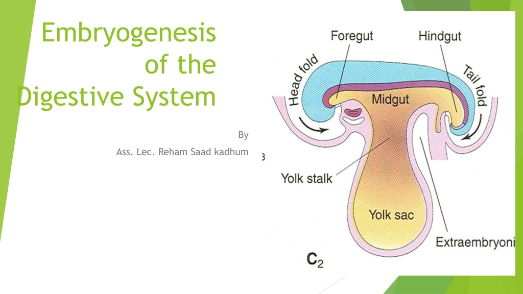
Formation of the gut tube during embryogenesis involves cephalocaudal and lateral folding of the embryo, resulting in the development of the foregut, midgut, and hindgut. The yolk sac and the allantois play key roles in the formation of the primitive gut. As the embryo develops, the vitelline duct and connecting stalk fuse with the umbilical cord. The fate of the gut tube mesentery includes the differentiation of ventral and dorsal mesogastrium, giving rise to structures like the liver, falciform ligament, lesser omentum, and more.

Download Presentation

Please find below an Image/Link to download the presentation.
The content on the website is provided AS IS for your information and personal use only. It may not be sold, licensed, or shared on other websites without obtaining consent from the author. Download presentation by click this link. If you encounter any issues during the download, it is possible that the publisher has removed the file from their server.
E N D
Presentation Transcript
Embryogenesis of the Digestive System By Ass. Lec. Reham Saad kadhum
Formation of the gut tube A result cephalocaudal and lateral folding of embryo. the endoderm-lined yolk sac cavity formed the primitive gut. Two other endoderm-lined cavity, the yolk sac and the allantois, remain outside the embryo. The primitive gut tube formed of three parts; the foregut,the midgut, and the hindgut.
Formation of the gut tube The anterior end of the foregut is closed by oral plate (or buccopharyngeal membrane ). The posterior end of the hindgut is closed by cloacal membrane. Midgut connected to the yolk sac by vitelline or omphalomesenteric duct.
Formation of the gut tube yolk sac is contribute to the ventral wall of the gut tube, the allantoisis fused with hindgut. end of the 4thweek, the vitelline duct and the connecting stalk will be fused with umbilical cord.
Fate of the gut tube mesentery: 1.The foregut tube by the ventral and dorsal mesogastrium. 2.The midgut and hindgut by dorsal mesogastriumonly. Elongated gut tube lead to absorbanceand disappearanceof some of these mesogastrium by a process called zygosis.
Fate of the gut tube mesentery: 1.Derivatives of the ventral mesogastrium of the foregut: This mesogastrium is divided by the growth of the liver inside it into two parts; a)The ventral mesogastrium: ventral to the liver------falciform ligament and other peritoneal ligaments of the liver. b)The ventral mesogastrium: dorsal to the liver-----lesser omentum.
Fate of the gut tube mesentery: 2.Derivatives of the dorsal mesogastrium of the foregut: mesogastrium divided by the growth of the spleen inside it into two parts: a.The dorsal mesogastrium ventral to the spleen -----greater omentum, the gastrosplenic ligament, and the gastrophrenic ligaments of the peritoneum. b.The dorsal mesogastrium dorsal to the spleen -------lienorenal peritoneal ligaments.
Fate of the gut tube mesentery These will give rise to all the peritoneal ligaments and mesenteries apart from those described above 3.Derivatives of the dorsal mesogastrium of the midgut and hindgut: Dorsal mesentery: suspended the caudal part of the foregut, the midgut, and a major part of the hindgut from the abdominal wall . Forms : Dorsal mesogastrium(greater omentum). Dorsal mesoduodenum Mesentery proper Dorsal mesocolon :
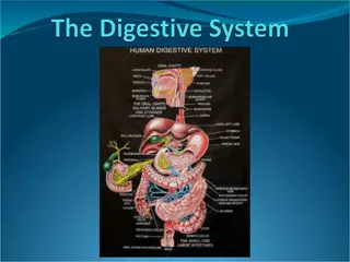
Blood supply for the derivatives of the gut tube: theforegut tube supplied by celiac trunk, the midgut tube supplied by superior mesenteric artery. the hindgut tube supplies by inferior mesenteric artery.
Derivatives of the foregut tube : 1.The Esophagus: The respiratory or tracheobronchial diverticulum appears at a ventral wall of the foregut at the border with the pharyngeal gut. This tracheobronchial diverticulum separates from the esophagus by tracheoesophageal septum. the esophagus is formed after division foregut tube into two parts by the tracheoesophageal septum. The ventral part forms the tracheobronchial diverticulum, The dorsal part forms the esophagus.
Clinical Correlates Esophageal Abnormalities: a)Esophageal atresia (with or without tracheobronchial fistula): The upper part of the esophagus doesn t connect with the lower esophagus and stomach. It usually ends in a pouch, which means food can t reach the stomach. It often happens along with another birth defect called a tracheoesophageal fistula, which is a connection between the lower part of the esophagus and the windpipe (trachea). b)Polyhydramnios: atresia of the esophagus prevents normal passage of amniotic fluid into the intestinal tract, resulting in accumulation of excess fluid in the amniotic sac is called polyhydramnios. c)Esophageal stenosis: the lumen of the esophagus may narrow, producing esophageal stenosis, usually in the lower third. d)Congenital hiatal hernia: the esophagus fails to lengthen sufficiently, and stomach is pulled up into the esophageal hiatus through the diaphragm.
Derivatives of the foregut tube 2. Stomach The stomach appears as a fusiform dilation of the foregut caudal to esophagus during the 4 th week of development. It rotates 90 degrees clockwise around its longitudinal axis, causing its left side to face anteriorly surface deviates to the right side to face posteriorly. Also the stomach rotates around its anteroposterior axis so that its caudal end moves upward and to the right , while its cephalic end moves downward and to the left.
Clinical Correlates Stomach Abnormalities: 1.Pyloric stenosis: Pyloric stenosis is narrowing of the opening from the stomach to the first part of the small intestine known as the duodenum. Due to enlargement (hypertrophy) of the circular muscle surrounding this opening , causes severe projectile vomiting. It occurs in the first few months of life. 2.Other malformation of the stomach, such as duplication of the stomach and prepyloric septum are rare.
Derivatives of the foregut tube : 3. upper duodenum (down to the level of the bile duct opening): This part of the duodenum which is derived from the foregut rotates with the stomach to the right forming its C shaped configuration . It becomes retroperitoneal due to the absorption of the mesentery .
Derivatives of the foregut tube 4. The liver: It develops as a diverticulum from the caudal end of the foregut during the 3rdweek of development, It elongates into the mesoderm of the septum transversum. A ventral branch projects from the liver bud forming the gall bladder and the cystic duct. The liver bud branches profusely around the vitellineand umbilical veins forming the liver parenchyma. Other liver cells (other than the hepatocytes) are derived from the mesoderm of the septum transversum.
Clinical Correlates liver Abnormalities: 1.Atresia of the gall bladder and/or the bile ducts (intra or extra hepatic ducts). 2.Double gall bladder. 3.Gall bladder with mesentery. 4.accessory hepatic duct.
Derivatives of the foregut tube 5.Pancreas: It is derived from the two buds; a dorsal and ventralbuds. These buds are located inside the dorsal and ventral mesogastrium respectively. When the duodenum rotates, the ventral pancreas rotates dorsallyon the dorsal pancreas to forms the lower part of the head of the pancreas and the uncinate process. The duct of the dorsal pancreas forms the main pancreatic duct, while the duct of the ventral bud forms the accessory pancreatic bud.
Clinical Correlates Pancreas Abnormalities: 1.Annular pancreas; the ventral pancreas divides into two parts, one of these rotates to dorsaland on the right side of the duodenum and the other part rotates dorsaland on the left side of the duodenum and so surrounding the duodenum and may causes duodenal obstruction. 2.Accessory pancreatic tissues that may be found in the stomach or in other abnormal sites.
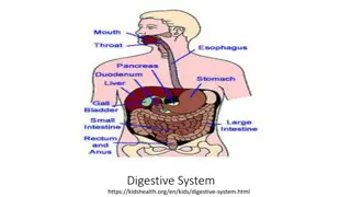
Derivatives of the midgut .1.the lower 2/3 of the duodenum below the opening of the bile duct. 2.the jejunum and the ileum. 3.the cecum,appendix, ascending colon, and right 2/3 of the transverse colon. The primary intestine projects out of the abdomen into the umbilical cord during the 6thweeks of development to form the physiological umbilical hernia.
Derivatives of the midgut This hernia returns into the abdomen during the 10thweek of gestation. The primary intestine rotates 270 degrees in a counter clockwise direction around its anteroposterior axis (which is the axis of the superior mesenteric artery) so that the cecum, the ascendingand transverse colon deviates up ward and to the right. The mesentery of the duodenum, cecum and ascending colon is absorbed by zygosis.
Clinical Correlates Abnormalities: 1.Persistent vitelline duct; forming the Meckel's diverticulum at the anti mesenteric border of the ileum. 2.Vitelline cyst; that may be twisted causing strangulation and gangrene. 3.Patent vitelline duct forming umbilical fistula with fecal discharge from the intestine out of the umbilicus. 4.Omphalocele;the physiological intestinal hernia is not returned into the abdominal cavity.
Clinical correlates : Abnormality Congenital umbilical hernia; by the absence of the skin and muscles of the anterior abdominal wall. 5.Abnormal rotation of the intestine; that may cause left sided colon or there may be reversed rotation of the intestine. 6.Duplication of the intestine. 7.atresia or stenosis of the intestine.
Derivatives of the hindgut: 1.The left 1/3 of the transverse colon. 2.Descending colon. 3.Sigmoid colon. 4.The rectum. 5.Upper part of the anal canal. The lower part of the anal canal is derived from the ectodermal anal pit or called the proctodeum. The junction between the endodermal and ectodermal parts of the anal canal is demarcated by the cloacal membrane.
Clinical Correlates Abnormalities ; Imperforated anus; occurring due to failure of cloacal membrane to rupture. 2.Rectal atresia. 3.Anal stenosis. 4.Rectal fistula between the rectum and the vagina or the urinary bladder, the perineum, or the urethra.