Pathophysiology of Cardiac Failure and Adaptive Mechanisms of the Heart

Cardiac failure is a condition where the heart is unable to meet tissue metabolic needs despite normal or increased venous return. Causes include decreased contractility, coronary blood flow, damaged valves, and more. Normal resting cardiac output is 5 Lt/mts, with adaptive mechanisms like the Frank-Starling mechanism and ventricular hypertrophy responding to increased load. Preload and myocardial contractility play crucial roles in cardiac function.

Download Presentation

Please find below an Image/Link to download the presentation.
The content on the website is provided AS IS for your information and personal use only. It may not be sold, licensed, or shared on other websites without obtaining consent from the author. Download presentation by click this link. If you encounter any issues during the download, it is possible that the publisher has removed the file from their server.
E N D
Presentation Transcript
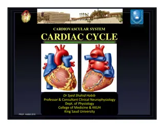
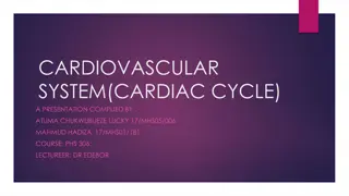
Pathophysiology of Cardiac Failure Professor Narsingh Verma
Introduction and Definition It is the pathophysiological process in which the heart as a pump is unable to meet the metabolic requirements of the tissue for oxygen and substrates despite the venous return to heart is either normal or increased Failure of Heart to function adequately Pumping ability is inadequate May be due to impaired contractile power Ventricular failure Right, left or both Acute as in Myocardial Infarction Chronic
Common causes Inability of Heart to Pump Decreased Contractility of Myocardium Diminished Coronary blood flow Damaged Heart Valves External Pressure around the heart Vitamin B Deficiency Primary cordiac muscle Diseases
Normal Resting cardiac output is 5 Lt/mts Heavy exercise it can go up to 25lt/mts Large reserve capacity In early cardiac failure this reserve is utilized = compensation Decompensation leads to clinical manifestations
Adaptive mechanisms of the heart to increased load Frank - Starling mechanism Ventricular hypertrophy Increased mass of contractile elements strength of contraction Increased sympathetic adrenergic activity Increased HR, increased contractility Incresed activity of R A A system
Preload Stretching the myocardial fibers during diastole by increasing end- diastolic volume force of contraction during systole = Starling s law preload = diastolic muscle sarcomere length leading to increased tension in muscle before its contraction - venous return to the heart is important volume is influenced end-diastolic - stretching of the sarcomere maximises the number of actin-myosin bridges responsible for development of force - optimal sarcomere length 2.2 m
Myocardial contractility Contractility of myocardium Changes in ability of myocardium to develop force by contraction that occurs independently on changes in myocardial fibre length Mechanisms involved in changes of contractility amount of created cross-bridges in the sarcomere by of Ca ++ i - catecholamines Ca++ i - inotropic drugs contractility Ca++ i contractility contractility shifting the entire ventricular function curve upward and to the left contractility shifting the entire ventricular function curve (hypoxia, acidosis) downward and to the right
Afterload It is expressed as tension which must be developed in the wall of ventricles during systole to open the semilunar valves and eject blood to aorta/pulmunary artery Laplace law: intraventricular pressure x radius of ventricle wall tension = -------------------------------------------------------- 2 x ventricular wall thickness afterload:due to - elevation of arterial resistance - ventricular size - intrathoracic pressure (loss of myocard) afterload:due to - arterial resistance - myocardial hypertrophy - ventricular size
Definition of the terms Myocardial failure = abnormalities reside in the myocardium and lead to inability of myocardium to fulfill its function Circulatory failure = any abnormality of the circulation responsible for the inadequacy in body tissue perfusion, e.g. decreased blood volume, changes of vascular tone, heart function disorders Congestive heart failure = clinical syndrome which is developed due to accumulation of the blood in front of the left or right parts of the heart
General pathomechanisms involved in heart failure development Cardiac mechanical dysfunction can develop as a consequence in preload, contractility and afterload disorders Disorders of preload preload length of sarcomere is more than optimal strength of contraction preload length of sarcomere is well below the optimal strength of contraction
Important:failing ventricle requires higher end-diastolic volume to achieve the same CO that normal ventricle achieves with lower ventricular volumes Disorders of contractility In the most forms of heart failure the contractility of myocardium is decreased (ischemia, hypoxia, acidosis, inflammation, toxins, metabolic disorders... ) Disorders of afterload due to: fluid retention in the body increased blood volume arterial resistance valvular heart diseases ( stenosis )
Characteristic features of systolic dysfunction (systolic failure) ventricular dilatation reducing ventricular contractility (either generalized or localized) diminished ejection fraction (i.e. that fraction of end-diastolic blood volume ejected from the ventricle during each systolic contraction less then 45%) in failing hearts, the LV end-diastolic volume (or pressure) may increse as the stroke volume (or CO) decreases
Characteristic features of diastolic dysfunctions (diastolic failure) ventricular cavity size is normal or smaller than normal myocardial contractility is normal or hyperdynamic ejection fraction is normal (>50%) or supranormal ventricle is usually hypertrophied ventricle is filling slowly in early diastole (during the period of passive filling) end-diastolic ventricular pressure is increased
Causes of heart pump failure A. MECHANICAL ABNORMALITIES 1. Increased pressure load central (aortic stenosis, aortic coarctation...) peripheral (systemic hypertension) 2. Increased volume load valvular regurgitation hypervolemia 3. Obstruction to ventricular filling valvular stenosis pericardial restriction
B. MYOCARDIAL DAMAGE 1. Primary a) cardiomyopathy b) myocarditis c) toxicity (e.g. alcohol) d) metabolic abnormalities (e.g. hyperthyreoidism) 2. Secondary a) oxygen deprivation (e.g. coronary heart disease) b) inflammation (e.g. due to increased metabolic demands) c) chronic obstructive lung disease
Pathomechanisms involved in heart failure A. Pathomechanisms involved in myocardial failure 1. Damage of cardiomyocytes contractility, compliance Consequences: defect in ATP production and utilisation changes in contractile proteins uncoupling of excitation contraction process number of cardiomyocytes impairment relaxation of cardiomyocytes with decrease compliance of myocardium impaired of sympato-adrenal system (SAS) 1-adrenergic receptors on the surface of cardiomycytes number of
2. Changes of neurohumoral control of the heart function Physiology: SNS contractility HR activity of physiologic pacemakers Mechanism: sympathetic activity Ca ++ i cAMP contractility sympathetic activity of parasympathetic system on the heart influence Pathophysiology:normal neurohumoral control is changed and creation of pathologic neurohumoral mechanisms are present
Nitric oxyde Bradykinin Endothelin Pro-proliferative effects Anti-proliferative effects
Chronic heart failure (CHF) is characterized by an imbalance of neurohumoral adaptive mechanisms with a net results of excessive vasoconstriction and salt and water retention Catecholamines :- concentration in blood : - norepinephrin 2-3x higher at the rest than in healthy subjects - circulating norepinephrin is increased much more during equal load in patients suffering from CHF than in healthy subject - number of beta 1 adrenergic receptors sensitivity of cardiomyocytes to catecholamines contractility System rennin angiotensin aldosteron heart failure CO kidney perfusion stim. of RAA system
Important: Catecholamines and system RAA = compensatory mechanisms heart function and arterialBP The role of angiotensin II in development of heart failure vasoconstriction (mainly in resistant vesels) retention of Na blood volume releasing of arginin vasopresin peptide (AVP antidiuretic hormon) from neurohypophysis
facilitation of norepinephrine release from sympathetic nerve endings sensitivity of vessel wall to norepinephrine mitogenic effect on smooth muscles in vessels and on cardiomyocytes in the heart hypertrophy mitogenic effect on fibrocytes in vessel wall and in myocardium constriction of vas efferens (in glomerulus) sensation of thirst secretion of aldosteron from adrenal gland mesangial conctraction glomerular filtration rate
Pathogenesis of heart failure Index event primary cause of heart damage Secondary damage remodeling Adrenergic, RAA, cytokine systems are involved in the remodeling Douglas L. Mann, 2004
Pathophysiology of diastolic heart failure systolic heart failure = failure of ejecting function of the heart diastolic heart failure = failure of filling the ventricles, resistance to filling of ventricles Diastolic failure is a widely recognized clinical entity But, which of the cardiac cycle is real diastole ?
Definition of diastolic heart failure It is pathophysiological process characterized by symptoms and signs of congestive heart failure, which is caused by increased filling resistance of ventricles and increased intraventricular diastolic pressure Primary diastolic heart failure - no signs and symptoms of systolic dysfunction is present - ! up to 40% of patients suffering from heart failure! Secondary diastolic heart failure - diastolic dysfunction is the consequence of primary systolic dysfunction
Main causes and pathomechanisms of diastolic heart failure 1. structural disorders passive chamber stiffness a) intramyocardial e.g. myocardial fibrosis, amyloidosis, hypertrophy, myocardial ischemia... b) extramyocardial e.g. constrictive pericarditis 2. functional disorders myocardial ischemia, advanced hypertrophy of ventricles, failing myocardium, asynchrony in heart ventricle functions relaxation of chambers e. g.
Causes and mechanism participating on impaired ventricular relaxation a)physiological changes in chamber relaxation due to: prolonged ventricular contraction Relaxation of ventricles is not impaired ! b) pathological changes in chamber relaxation due to: Impaired relaxation process delayed relaxation (retarded) incomplete (slowed) relaxation
Consequences of impaired ventricular relaxation - filling of ventricles ismore dependent on diastasis and on the systole of atrias than in healthy subjects Symptoms and signs: exercise intolerance = early sign of diastolic failure coronary blood flow during diastole Causes and mechanisms involved in development of ventricular stiffness ventricular compliance= passive property of ventricle Source of compliance: cardiomyocytes and other types of cells in the heart tissue to stretching
Ventricular compliance is caused by structural abnormalities localized in myocardium and in extramyocardial tissue a) Intramyocardial causes : myocardial fibrosis, hypertrophy of ventricular wall, restrictive cardiomyopathy b. Extramyocardial causes : constrictive pericarditis The role of myocardial remodelling in genesis of heart failure adaptive remodelling of the heart pathologic remodelling of the heart
Main causes and mechanisms involved in pathological remodelation of the heart 1.Increased amount and size of myocytes = hypertrophy Due to:- volume and/or pressure load (excentric, concentric hypertrophy) - hormonal stimulation of cardiomyocytes by norepinephrine, angiotenzine II, endothelin... 2. Increased % ofnon-myocytic cellsin myocardium and their influence on structure and function of heart a.endothelial cells endothelins : mitogenic ability stimulation growth of smooth muscle cells of vessels, fibroblasts b.fibroblasts - production of kolagens
Left ventricular Failure Primary Ventricular Dysfunction Arterial Hypertension Inadequate left ventricular pumping ------Building up high pressure in left atrium ------back pressure transmitted to pulmonary veins and capillaries ------pulmonary congestion ------may produce Crepitation -- -can be heard by auscultation-------may lead to pulmonary edema---- symptoms of Cough and Dyspnea ---this congestion is more in lying down posture than when sitting up ---breathing difficulty is also more during lying down position ----Orthopnea Decreased blood flow to tissues ----Tissue Hypoxia
Right Ventricular failure Primary ventricular dysfunction More commonly secondary to an overload which may result from Mitral stenosis or Parenchymal lung disease ( Emphysema or Fibrosis) Cor pulmonale Right Ventricular Failure ------Back Pressure in the Right Atrium and Systemic Veins -----Venous congestion ----Increased Jugular venous pressure ----Pedal Edema, Ascites and Enlarged Liver
Biventricular Cardiac Failure All left and Right ventricular failure ultimately becomes Biventricular Left ventricular failure ------------Increase in left atrial Pressure ---------- --------------------------Increase in pulmonary capillary pressure ------------ --------Increase in Pulmonary arterial pressure ------------------------------- --------------Right ventricular hypertrophy ---------------------------Right Ventricular Failure
High Output cardiac Failure Paradoxical Situation Left to right Shunt tetralogy of Fallot -----Blood flows from the left side to right side --------out put of right ventricle is high ------- eventually right ventricle fails Hyperthyroidism High metabolic rate -----increased blood flow to meet the enhanced requirements-----High Cardiac output -----Heart failure Severe Anemia
In left ventricular failure there are more symptoms but only sign is fine crepitation Right ventricular failure patient has raised jugular venous pressure, Prominent Jugular veins, hepatomegaly and edema but may not have any symptoms


 is characterized by an")