Understanding Shock: Etiology, Pathophysiology, Diagnosis, and Management Priorities

Shock, a condition where blood circulation fails to deliver sufficient oxygen, is a common cause of death in surgical patients. Recognizing shock early is crucial to prevent irreversible tissue damage and organ failure. It is vital to understand the various aspects such as etiology, pathophysiology, diagnosis, and management priorities associated with shock to provide timely and effective treatment.

Download Presentation

Please find below an Image/Link to download the presentation.
The content on the website is provided AS IS for your information and personal use only. It may not be sold, licensed, or shared on other websites without obtaining consent from the author. Download presentation by click this link. If you encounter any issues during the download, it is possible that the publisher has removed the file from their server.
E N D
Presentation Transcript
Shock HAMAD ALQAHTANI Professor of Surgery Consultant Hepatobiliary Surgeon College of Medicine King Saud University
Definition Definition Shock exists when circulating blood has failed to deliver the required oxygen to meet the cell s metabolic requirements.
Introduction Introduction Shock is the commonest cause of death in surgical patients. 1. Early and rapidly: Severe profound shock state. 2. Late: Ischemia, reperfusion, and organs failure. It is important to understand the following points in shock 1. Etiology 2. Pathophysiology 3. Diagnosis 4. Management priorities of shock. Early recognition and treatment of shock is needed to prevent irreversible tissue damage and organs failure.
Shock state does not mean hypotension. Hypotension is one of the latest signs of shock. Young healthy patients are able to maintain blood pressure until late stages of shock by increasing: 1. Stroke volume 2. Peripheral vasoconstriction.
Old patient decompensate earlier than the young patient. Beta-blockers and pacemaker can prevent the tachycardic response and the diagnosis of shock can be challenging in this situation.
consequences of delayed or untreated consequences of delayed or untreated shock shock 1. Reduced oxygen consumption 2. Anaerobic cell metabolism 3. Tissue acidosis 4. Cellular dysfunction 5. Multiple organ dysfunction 6. Death
Types of shock Types of shock (1) Hypovolemic shock It is the commonest type and the most treatable cause of shock in surgical practice. It is due to reduced intravascular fluid volume secondary to: Hemorrhagic causes 1. Trauma 2. Surgery 3. Gastrointestinal 4. Others
Non-hemorrhagic causes 1. 2. Excessive fluid loss due to vomiting, diarrhea, fistula, urinary loss (diabetes ketoacidosis). 3. Evaporation during prolonged surgery 4. Third spacing where the fluid is lost into the interstitial compartment (e.g. acute pancreatitis or burn). Poor fluid intake
Severity of hypovolemic shock Severity of hypovolemic shock The severity of hemorrhagic shock is classified according to the percentage of estimated blood volume loss.
Classification of Hypovolaemic Shock Classification of Hypovolaemic Shock Class I Class I Class II Class II Class III Class III Class IV Class IV Normal Normal Anxious Anxious Confused Confused Lethargic Lethargic CNS Symptoms CNS Symptoms < 750 < 750 750 750 1500 1500 1500 1500 2000 2000 > 2000 > 2000 Blood Loss (ml) Blood Loss (ml) < 15 % < 15 % 15 15 30% 30% 30 30 40% 40% > 40% > 40% Blood Loss Blood Loss (% EBV) (% EBV) < 100 < 100 > 100 > 100 > 120 > 120 > 140 > 140 Pulse Pulse Normal Normal Decreased Decreased Decreased Decreased Decreased Decreased Blood Pressure Blood Pressure 14 14 20 20 20 20 30 30 30 30 35 35 > 35 > 35 Respiratory Rate Respiratory Rate > 30 > 30 20 20 30 30 5 5 15 15 Negligible Negligible Urine Output Urine Output (ml/h) (ml/h)
(2) Septic shock It can be defined as sepsis-induced hypotension that persist despite adequate intravascular fluid resuscitation. It causes disturbances in oxygen delivery and consumption.
Sepsis usually arises from a localized bacterial focus of infection in the lungs, abdomen, pelvis, urinary tract, and skin. It is most commonly due to Gram-positive, followed by endotoxin-producing Gram-negative bacteria.
Infection in septic shock triggers a cytokine-mediated proinflammatory response which results in: 1. Peripheral vasodilation 2. Redistribution of blood flow 3. Endothelial cell activation 4. Increased vascular permeability 5. Formation of microthrombi within the microcirculation
Typically cardiac output increases in septic shock in compensation for the peripheral vasodilation. Microcirculatory dysfunction in septic shock impairs the delivery of oxygen to the cells, despite hyperdynamic circulation with a global increase in oxygen delivery.
The late stage of septic shock characterized by hypotension due to: 1. Vasodilatation 2. Loss of intravascular fluid to the interstitial compartment (third spacing resulting in edema) 3. Myocardial depression
(3) Cardiogenic Shock This occurs due to heart failure (pump failure) to maintain the cardiac output that meets the metabolic requirements of the body. Causes of cardiogenic shock: 1. Arrhythmias 2. Cardiomyopathy 3. Myocardial infarction 4. Valvular heart disease 5. Blunt myocardial injury
6. Myocardial depression (e.g. humoral agents during sepsis or drugs) 7. Cardiac tamponade 8. Massive pulmonary embolism 9. Tension pneumothorax
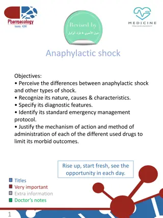
(4) Anaphylactic Shock This is a severe systemic hypersensitivity reaction following exposure to an allergen which triggers the release of vasoactive mediators (prostaglandins, histamine, and kinins) from basophils and mast cells. This systemic reaction cause cardiovascular changes: 1. Vasodilation 2. Intravascular volume redistribution 3. Capillary leak 4. Reduction in cardiac output shock by the following
Common causes of anaphylaxis in surgical practice: 1. Drugs (e.g. -lactam antibiotics) 2. Colloid solutions (e.g. dextrans) 3. Radiological contrast media (intravenous injection of contrast) 4. Latex (e.g. latex medical gloves)
(5) Neurogenic Shock This is due to loss of sympathetic tone in the vascular smooth muscle. This occurs most commonly due to injury to the cervical or thoracic spinal cord and results in: 1. Profound vasodilation 2. Fall in systemic vascular resistance 3. Warm, flushed skin due to vasodilation 4. Bradycardia 5. Hypotension 6. Respiratory arrest (injury above the 3rd cervical vertebra)
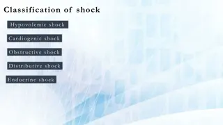
The terminology used in the Classification of Shock Obstructive Shock This term is used to indicates the reduction in the preload and cardiac filling. 1. Massive pulmonary embolism 2. Air embolism 3. Tension pneumothorax 4. Cardiac tamponade
Distributive Shock This response is characterized by inadequate tissue perfusion, vasodilatation, hypotension, low systemic vascular resistance, and low afterload. All results in the high cardiac output. 1. Neurogenic 2. Septic 3. Anaphylactic shock
Pathophysiology of shock Pathophysiology of shock Cells 1. Deprivation of cells from oxygen 2. Switch of the cell s metabolism from aerobic to anaerobic 3. Lactic acidosis
4. Failure of sodium/potassium pump in the cell membrane and intracellular organelle failed which results in two major events: Intracellular release of autodigestive enzymes from lysosomes resulting in cell lysis and death. The release of intracellular contents, including the potassium into the systemic circulation.
Microcirculation Progression of hypoxia, ischemia, and acidosis will activate the immune system (neutrophils and complements) and coagulation system. 1. Release of oxygen free radicles and cytokines 2. Injury to the endothelial cells 3. Further activate the immune and complement systems 4. Loss of the integrity of endothelial cells and becomes leaky . 5. Fluid loss through the spaces between the endothelial cells will cause tissue edema and exacerbation of cellular hypoxia.
Systems/Organs Adequate perfusion and oxygen delivery to vital organs (brain, heart, skeletal muscle) will compensatory mechanisms have failed. be compromised when In severe, prolonged or uncorrected shock ( decompensated shock), the clinical manifestations of organ hypoperfusion become apparent due to prolonged severe hypoxia and acidosis which may result in organ dysfunction and failure.
Nervous System 1. Restlessness 2. Confusion 3. Stupor 4. Coma 5. Encephalopathy and/or delirium are common in sepsis or septic shock
Cardiovascular System 1. Reduced preload and afterload baroreceptor responses (increased sympathetic response and release of catecholamines) vasoconstriction (except in septic shock). compensatory tachycardia and systemic 2. Decrease diastolic pressure reduced coronary blood flow.
3. Decrease myocardial oxygen delivery may result in: Myocardial ischemia Decreased myocardial contractility Decreased cardiac output. 4. Acidosis, electrolyte disturbances and hypoxia arrhythmias 5. Widespread endothelial cell activation dysfunction. microcirculatory
Respiratory System 1. The increased sympathetic response and metabolic acidosis result in: Tachypnea Increased minute ventilation to increase carbon dioxide elimination (compensatory respiratory alkalosis). 2. Increased ventilation/perfusion mismatch and shunting, which will result in hypoxia
3. Pulmonary edema (common in cardiogenic shock) with hypoxia 4. Acute lung injury and acute respiratory distress syndrome with hypoxia
Gastrointestinal Tract 1. Splanchnic hypoperfusion will lead to the breakdown of the gut mucosal barrier 2. Translocation of bacteria/bacterial wall contents into the bloodstream will lead to the systemic inflammatory response syndrome and sepsis
3. Acute ischaemic hepatitis (in the severe case may result in prolonged prothrombin time, hypoglycemia and very high transaminases ALT and AST) 4. Stress ulceration and gastroduodenal bleeding 5. Decreased motility and impaired absorption
Renal System 1. Activation of the renin-angiotensin system: Vasoconstriction Sodium and water reabsorption in the kidney. 2. Oliguria (< 0.5 ml/kg/h urine) 3. Anuria 4. Acute renal failure (Acute tubular necrosis) with high urea and creatinine, hyperkalemia and metabolic acidosis.
Endocrine System 1. Adrenal activation with release of catecholamines 2. Renin-angiotensin-aldosterone increase reabsorption of sodium followed by water in the distal renal tubules and collecting ducts of the kidneys. system activation, which
Release of vasopressin (antidiuretic hormone-ADH) from the hypothalamus Vasoconstriction Water resorption in the renal collecting tubules. Cortisol release from adrenal glands Sodium and water resorption Sensitization of the cells to catecholamines.
Ischemia-Reperfusion Effect of Shock 1. Hypoperfusion and hypoxia 2. Damage to the cells and organs 3. Restoration of normal organ perfusion cause further damage 4. Acidosis and hyperkalemia 5. Myocardial depression 6. Vasodilatation with further aggravation of hypotension. 7. Neutrophils, complements and microthrombi will be flushed back into circulation after reperfusion 8. Endothelial injury in the lung, kidneys and other organs. 9. Acute renal, and lung injury, other organ failure, and death. 10. This ischemia-reperfusion effects can only be attenuated by reducing the extent and duration of hypoperfusion.
Clinical features of shock Initially, the endocrine and cardiovascular compensatory responses reduce the blood flow to non-essential organs (skin, muscles, gastrointestinal tract) to maintain the blood flow to the kidneys, lungs, and brain. During this stage, there is metabolic acidosis and activation of humoral and cellular elements in the non-perfused organs.
If the shock is inadequately treated, the compensation stage will result in decompensation with: 1. Profound hypotension 2. Tachycardia 3. Hypoperfusion of organs, This result in: 1. Impaired level of consciousness 2. Oliguria 3. Anuria 4. Increased in lactic acid 5. Ultimately organ failure and death. Tachycardia may not be a manifestation of shock in some patients, such as the patient on beta-blocker or implanted cardiac pacemaker. These patients are unable during shock to mount a tachycardia.
Mild Shock 1. Tachycardia 2. Tachypnea 3. Mild drop in the urine output 4. Mild anxiety 5. Blood pressure is maintained despite 6. Decreased pulse pressure 7. Peripheries are cool 8. Sweaty with prolonged capillary filling time Patients with septic shock will have flushed, warm peripheries, with briskly capillary refilling despite profound shock.
Moderate Shock 1. Tachycardia 2. Drop in the blood pressure 3. Mildly confused 4. Drowsy. 5. Urine output below 0.5 ml / kg/hour.
Severe Shock 1. Profound tachycardia 2. Profound hypotension 3. Unconsciousness 4. Labored respiration 5. Urine output drop to zero (anuria)
Management of shock Hypovolemic Shock Airway, breathing, and circulation (ABC) Oxygenation Fluid resuscitation (crystalloid or colloid). Urinary catheterization is required to monitor the urine output. Blood transfusion.
Control of hemorrhage Analgesia Inotropes Admission Massive transfusion can cause: Hypothermia Hypocalcemia Hyper-or hypokalaemia Coagulopathy
Septic Shock The most important aspect of septic shock is early recognition, which requires a high index of suspicion, with a detailed history and physical examination to identify potential sources of infection and signs of organ dysfunction.
Septic shock is associated with relative and absolute hypovolemia as a result of: 1. Profound vasodilation 2. Fluid extravasation from the intravascular space to the interstitial space (third spacing). The two main principles guiding the management of septic shock are: 1. The identification and treatment of underlying infection 2. Early optimization of tissue oxygen delivery.
1. Airway, breathing, and circulation (ABC) 2. Fluid (crystalloid or colloid) resuscitation 3. Oxygenation. 4. Vasopressor 5. Intravenous hydrocortisone
6. Treatment of infection Intravenous antibiotics must be administered as soon as possible. It should cover all likely pathogens (bacterial and/or fungal). It usually involves the use of empirical broad-spectrum antibiotics to be changed according to the microbiological investigations (gram stain & culture and sensitivity). Peripheral blood culture should be taken prior to the antibiotics administration.
Targeted imaging, including chest x-ray, ultrasound, computed tomography may be required to localize the site of the focus of infection. Adequate source control which includes removal of infected devices abscess drainage and debridement of infected tissue Interventions to prevent ongoing microbial contamination such as biliary drainage or repair of a perforated viscus. It should be performed immediately after initial resuscitation and should be achieved with the minimum physiological disturbance. Percutaneous or endoscopic interventions are preferable to open surgery where possible.
Cardiogenic Shock Acute anterior commonest cause of cardiogenic shock. The major disturbances in cardiogenic shock are a reduced cardiac output with a compensatory increase in systemic vascular resistance. myocardial infarction is the


 Septic shock")


 Cardiogenic Shock")

 Anaphylactic Shock")

 Neurogenic Shock")


")


 from the")


")