Understanding the Cardiac Cycle: A Comprehensive Overview

The cardiac cycle, involving systole and diastole processes, consists of arterial and ventricular events essential for heart function. Arterial events include systole and diastole, while ventricular events encompass isometric contraction, ejection period, prodiastole, isometric relaxation, rapid filling, slow filling, and last rapid filling. Each event plays a crucial role in maintaining proper blood flow through the heart.

Download Presentation

Please find below an Image/Link to download the presentation.
The content on the website is provided AS IS for your information and personal use only. It may not be sold, licensed, or shared on other websites without obtaining consent from the author. Download presentation by click this link. If you encounter any issues during the download, it is possible that the publisher has removed the file from their server.
E N D
Presentation Transcript
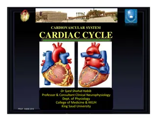
CARDIOVASCULAR SYSTEM(CARDIAC CYCLE) A PRESENTATION COMPLIED BY : ATUMA CHUKWUBUEZE LUCKY 17/MHS05/006 MAHMUD HADIZA 17/MHS01/181 COURSE: PHS 306: LECTUREER: DR EDEBOR
CARDIAC CYCLE Cardiac cycle is the series of event that takes place within one heart beat. It involves to processes namely: Systole: which is contraction of cardiac muscles Diastole: which is relaxation of cardiac muscles The cardiac cycle has two events Arterial event Ventricular event
ARTERIAL EVENTS Arterial Systole: It is also called late rapid filling or presystolic. It is when the atrium contract to remove the final 10% of blood in it to the ventricle. Hence about 90% of ventricular filling has taken place and simultaneously, the ventricular diastole takes place. Arterial Diastole: It is when the atrium relax and simultaneously, the ventricular systole. The atrium is filled with blood at this point.
VENTRICULAR EVENT(SYSTOLE) Isometric Contraction: It is also called isovolumetric contraction. It is when the pressure in the ventricle starts building up hence, closing the atrioventricular valve which is the first heart sound and the ventricle forms a close chamber. Simultaneously, the atrium undergoes relaxation. Ejection Period: This is when blood flows to the pulmonary and aortic trunks after opening of the semilunar valve due to increased pressure in the ventricle. The ejection is at first fast and reduce with time. The atrium is still relaxed and receive blood. Not all the blood is ejected from the heart some are left and it is call End Systolic Volume which is about 60-80ml.
VENTRICULAR EVENT (DIASTOLE) Prodiastole: This is the period that prepare the ventricle for diastole by reduction of pressure in the ventricle making the semilunar valve close which is the second heart beat. Isometric Relaxation: This is also called isovolumetric relaxation and it occurs when the pressure in the ventricle reduces hence, the atrioventricular valve and the semilunar valve are closed.
VENTRICULAR EVENT (DIASTOLE) Rapid Filling: This is when the ventricle is filled with blood due to opening of the atrioventricular valve about 70% of the blood in the atrium enters. Slow Filling: Only about 20% of blood enters the ventricle and it is also called Diastasis. Last rapid filling: Only about 10% of blood enters the ventricle due to atrial systole. And End Diastolic Volume is about 120- 130ml
PRESSURE AND VOLUME VENTRICULAR PRESSURE When there is contraction of the atrium the ventricle also forms the curve of the a wave. AORTIC PRESSURE When the ventricle is undergoing contraction, pressure build s up the aortic pressure is initiated. ARTERIAL PRESSURE When there is contraction of the atrium a wave is formed. When the ventricle is undergoing contraction, pressure build s up and forms c wave in the atrium. When the ventricle is undergoing contraction, pressure build s up and it depolarize to the region of aortic pressure about 80mmHg. When the semilunar valve opens the aortic curve reaches its peak. When the ventricle relax the atrial curve forms v wave. When the semilunar valve opens the ventricular curve reaches its peak. When the ventricle relax it reduces back to it s initial point 80mmHg When the ventricle relax it s curve repolarize back to initial point.


")
")
")
")