Understanding Dyspnea: Definition, Pathophysiology, and Causes

Dyspnea, or breathing discomfort, is a complex symptom with varying intensities and causes, including lung diseases, cardiac issues, anemia, obesity, and deconditioning. This article explores the definition, pathophysiology, and different approaches to understanding and managing dyspnea, highlighting the importance of recognizing both physical symptoms and emotional distress associated with this condition.

Download Presentation

Please find below an Image/Link to download the presentation.
The content on the website is provided AS IS for your information and personal use only. It may not be sold, licensed, or shared on other websites without obtaining consent from the author. Download presentation by click this link. If you encounter any issues during the download, it is possible that the publisher has removed the file from their server.
E N D
Presentation Transcript
SOB Khalid Al-Mobaireek
Introduction and Definition Pathophysiology Causes Approach cases
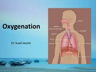
Dyspnea common symptom may be the primary manifestation of lung disease, cardiac, anemia, obesity, or deconditioning.
What is Dyspnea? Dyspnea: breathing discomfort Subjective experience of breathing discomfort that consists of qualitatively distinct sensations that vary in intensity ATS Consensus Statement 2012 Uncomfortable awareness of breathing Major problem: Lack of a valid, reliable measure of the degree of Dyspnea Physical symptoms define dyspnea intensity Emotional response determines degree of distress Dyspnea is complex multidimensional symptom
Dyspnea can be: acute develops over hours to chronic > 4-8 weeks. acute worsening of chronic breathlessness (eg, CF, heart failure).
Introduction and Definition Pathophysiology Causes Approach cases
receptors Chemoreceptors (peripherally and centrally located) mechanoreceptors (located in the airways, lungs and chest wall)
Chemoreceptors stimulated by changes in PCO 2 and PO 2 levels. Central chemorecepters are in the medulla. Peripheral chemoreceptors are in the aortic arch and carotid bodies
Mechanoreceptors located in the airways, lungs and the chest wall. Lungs: three main types: pulmonary stretch receptors: increased airway tension and lung volume (asthma related dyspnea) Irritant receptors: bronchial walls C-fibers: near alveoli in the pulmonary parenchyma, Chest wall receptors: respond to restricted chest wall movements.
PATHOPHYSIOLOGICAL FEATURES In normal:respiratory muscles work only during inspiration the diaphragm does most of the work. The work of inspiration is the sum of the work necessary to overcome: the elastic forces of the lung the tissue viscosity of the lung and chest wall airway resistance.
classic sign of dyspnea The accessory muscles of inspiration: the sternocleidomastoid anterior serratus external intercostal muscles Contraction of these muscles increases the ve intrathoracic pressure. The negative pressure draws in the soft tissues of the chest wall (retractions:suprasternal, infrasternal, intercostal, subcostal, and supraclavicular). Other way to maintain an adequate MV is to increase the RR(tachypnea). Nasal flaring MV= TV X RR
Expiration Little energy is expended during normal expiration. Relaxation of the diaphragm, elastic recoil of the lungs and chest wall, and compression of the lungs by the intraabdominal organs. In obstructive airway disease: more force is needed. In tachypnea: no time for elastic recoil to allow adequate exhalation between breaths. In either instance the accessory muscles of expiration are used: The abdominal recti muscles: force the abdominal contents against the diaphragm to compress the lungs the internal intercostal muscles contract to pull the ribs downward.
Respiratory cardiovascular neuromuscular systemic psychogenic Asthma/ CF/PCD/ bronchiectasis CHF/pul edema Rib #/ chest trauma anemia Hyperventilation syndrome Pneumonia/ Bronchiolitis arrhythmias Flail chest ARF Psychogenic dyspnea Pneumothorax Cyanotic spells Massive obesity Metabolic acidosis Vocal cord dysfunction Foreign Body Pul HPN kyphoscoliosis sepsis aspiration Valvular heart dis CNS/spinal cord disordes cirrohsis embolism Phrenic nerve palsy anaphylaxis Upper aw obs * Myopathy/ neuropathy fever * Foreign body (airway or esophagus), Croup, Epiglottitis, Retropharyngeal abscess, Enlarged tonsils or adenoids, tumors (vc, larynx, trachea, Mediastinum), Diphtheria, Bacterial tracheitis, Ingestion of caustic substance, Vascular ring, Tracheal laryngeal web, Bronchomalacia, choanal atresia
Introduction and Definition Pathophysiology Causes Approach cases
Approach Step 1: ABC (i.e is the patient stable?) a patient presenting with dyspnea must be evaluated for life threatening causes. acute in onset. FB, pneumothorax, tamponade. After these causes are ruled out, further evaluation can proceed.
History complete description of the dyspnea: Onset: sudden (eg, inhaled FB, pneumothorax) evolved over several hours (eg, asthma, diabetic ketoacidosis). duration the frequency of attacks of dyspnea Triggers severity (daily activities restriction. However, dyspnea sensation can be affected by the patient's anxiety level, previous experiences. Aggrivating factors: patient's position associated symptoms such as cough, wheezing, sputum, and pleuritic pain.
history of other known illnesses, allergies, illnesses in the family. social history: Deconditioning can be present in sedentary life style, smoking and environmental exposure. Medication effect can directly cause a sensation of dyspnea (e.g opioids) not compliant or recently stopped taking medications (such as lasix for fluid overload, or brochodilators).
Physical exam While taking the history you can also begin the inspection. vital signs including O2 sat. Does the patient have fever? Growth parameters Finger clubbing inspection of the upper airways What position is the patient in? (sitting forward, etc) Does the patient have stridor? Tracheal tug?
extrathoracic obstruction: Hoarsness (larynx)? Cough sound? Does the patient have signs of increased WOB (inter/subcostal retractions)? Consider intrathoracic respiratory causes. Does the patient have a wheeze? Does the wheeze respond to bronchodilators? Full cardiac and pulmonary exams.
Cardiac Does the child develop SOB during feeds or activity? Have poor weight gain? Does the anterior left hemithorax look more prominent? Does the patient have periorbitalema? Hepatomegaly? Does the patient have tachychardea more than tachypnea.
Important clinical clues Hypoxia: most worrisome endpoint of SOB. Central Cyanosis: Deoxygenated hemoglobin is at least 5g/100mL of blood, or that O2 sats have dropped below 85%. either severe respiratory disease or cyanotic CHD. Peripheral cyanosis: if without central cyanosis, is a sign of decreased peripheral (not necessarily hypoxia).
After basic evaluation, formulating a hypothesis: there is primary cardiac, pulmonary, or neurologic pathology multiple systems may be impacted simultaneously.
Lab ABG: can indicate the elevated CO 2 levels, acidosis Electrolyte abnormalities. complete blood count: anemia. EKG: heart rhythmsCXR: pneumonia, pulmonary edema, FB. CT scan of the chest: pulmonary embolism and a multitude of pulmonary and vascular diseases.
Echocardiography: strucure, pulmonary pressures, assessing systolic function Troponin testing to evaluate for myocardial ischemia BNP and nt-pro-BNP separate dyspnea due to CHF versus pulmonary causes. Cardiopulmonary exercise testing: in cases of exertional dyspnea, when cardiac or pulmonary disease is at early stages.