Anatomy and Physiology of the Throat: A Comprehensive Overview
The throat, comprising the mouth, pharynx, and associated structures, plays vital roles in digestion, communication, and breathing. Understanding the anatomy and physiology of the throat, from the oral cavity to the laryngopharynx, helps in comprehending functions like swallowing, sound production, and air passage. Explore the divisions of the oral cavity, parts of the pharynx, and their respective functions for a detailed insight into this essential anatomical region.

Download Presentation

Please find below an Image/Link to download the presentation.
The content on the website is provided AS IS for your information and personal use only. It may not be sold, licensed, or shared on other websites without obtaining consent from the author. Download presentation by click this link. If you encounter any issues during the download, it is possible that the publisher has removed the file from their server.
E N D
Presentation Transcript
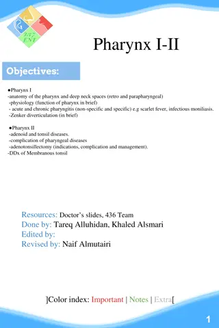
Anatomy and applied physiology of the throat . .
Mouth The is also known as the oral cavity. It has three major functions: Digestion receives food, transfer it the pharynx to preparing it for digestion in the stomach and small intestine. Communication modifies the sound produced in the larynx to create a range of sounds. Breathing acts as an air inlet in addition to the nasal cavity.
Anatomy The oral cavity lies between the oral fissure (anteriorly the opening between the lips), and the oropharyngeal isthmus (posteriorly the opening of the oropharynx .) The two divisions of the oral cavity are the vestibule, and the mouth cavity proper Vestibule It is the space between the lips/cheeks, and the gums/teeth Mouth proper The mouth proper lies posteriorly to the vestibule, the tongue fills a large proportion of the cavity of the mouth proper. The roof of the mouth proper consists of the hard and soft palates. The floor of the oral cavity consists of several structures :Muscular diaphragm comprised of the bilateral mylohyoid muscles, geniohyoid muscles, sublingual salivary glands and ducts
Pharynx It begins at the base of the skull, and ends at the inferior border of the cricoid cartilage (C6). The pharynx is comprised of three parts (superior to inferior): 1. Nasopharynx 2. Oropharynx 3. Laryngopharynx
Nasopharynx It is found between the base of the skull and the soft palate. It is continuous with the nasal cavity, and performs a respiratory function by conditioning inspired air and propagating it into the larynx. The posterosuperior wall of nasopharynx contains the adenoid tonsil , which enlarge between 3-8 years of age and then regress.
Oropharynx It is the middle part of the pharynx, located between the soft palate and the superior border of the epiglottis. It contains the following structures: 1. Posterior 1/3 of the tongue. 2. Lingual tonsils lymphoid tissue at the base of the tongue. 3. Palatine tonsils lymphoid tissue located in the tonsillar fossa (between the palatoglossal and palatopharyngeal 4. Superior constrictor muscle. Waldeyer s ring is the ring of lymphoid tissue in the naso- and oropharynx formed by the paired palatine tonsils, the adenoid tonsil, lingual tonsil and scattered lymph nodes. The oropharynx is involved in the voluntary and involuntary phases of swallowing
Laryngopharynx The most distal part of the pharynx, located between the superior border of the epiglottis and inferior border of the cricoid cartilage (C6). It is continuous inferiorly with the oesophagus. It is found posterior to the larynx and communicates with it via the laryngeal inlet, lateral to which one can find the piriform fossae. The laryngopharynx contains the middle and inferior pharyngeal constrictors
Muscles of pharynx There are two main groups of pharyngeal muscles; Circular 1. Superior pharyngeal constrictor 2. Middle pharyngeal constrictor . 3.Inferior pharyngeal constrictor All pharyngeal constrictors muscles are innervated by the vagus nerve (CN X) longitudinal muscles 1. Stylopharyngeus it is innervated by the glossopharyngeal nerve 2. Palatopharyngeus Innervated by the vagus nerve 3. Salpingopharyngeus Innervated by the vagus nerve
Blood supply of the pharynx Arterial supply to the pharynx is via branches of the external carotid artery: 1. Ascending pharyngeal artery 2. Branches of the facial artery 3. Branches of the lingual and maxillary arteries. Venous drainage is achieved by the pharyngeal venous plexus, which drains into the internal jugular vein
larynx The cartilaginous framework A. Unpaired cartilages 1. Epiglottis 2.The cricoid cartilage B. Paired cartilages 1. The arytenoids cartilages 2. The thyroid cartilages
Intrinsic muscles 1. Muscle that open the vocal cords Posterior cricoarytenoid muscle 2. Muscles that close the vocal cords A-Lateral cricoarytenoid muscle B- Interarytenoid musle C- Cricothyroid muscle 3. Muscle that increase the tension of the vocal cords Thyroarytenoid (vocalis) muscle
Arterial blood supply 1. Superior Laryngeal artery: branch of superior thyroid artery 2. Inferior Laryngeal artery: branch of inferior thyroid artery 3. Cricothyroid artery: branch of superior thyroid artery
Innervations Motor innervations by the recurrent laryngeal nerve that supplies all the intrinsic muscles of the larynx except the cricothyroid muscle which is supplied by the external laryngeal nerve which is branch of superior laryngeal nerve Sensory innervations of the larynx for the area above the vocal cords is supplied by the internal laryngeal branch of superior laryngeal nerve while the area below the vocal cords is supplied by the recurrent laryngeal nerve.
Functions of the larynx 1. Respiration. 2. Phonation (voice). 3. Protection of lower airways. 4. Fixation of chest