Understanding Dentin: Structure, Properties, and Functions
Dentin is a vital component of teeth, featuring various structural units such as dentinal tubules. This calcified tissue determines tooth shape, hardness, and radiolucency. It plays a significant role in supporting enamel, cementum, and pulp, with dentinal tubules housing odontoblast processes. Dentin's physical and chemical properties vary, contributing to its yellowish color, hardness, and mineral composition. Learn about its importance in dental health and function.

Download Presentation

Please find below an Image/Link to download the presentation.
The content on the website is provided AS IS for your information and personal use only. It may not be sold, licensed, or shared on other websites without obtaining consent from the author. Download presentation by click this link. If you encounter any issues during the download, it is possible that the publisher has removed the file from their server.
E N D
Presentation Transcript
Lec. Lec.8 8 Dentin Dentin Lecturer/Sura Haddad Msc in Histology Collage of Dentistry ,University of Basrah Department of Oral Diagnosis
Tips: Definition of the dentin Enumeration of the structure units of dentin Description of the structure units of dentin Description the types of incremental lines in dentin Definition of the interlobular dentin Enumeration and description of types of dentin
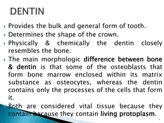
Dentin Dentin is a calcified tissue of the body, and along with enamel, cementum, and pulp is one of the four major components of teeth. It is usually covered by enamel on the crown and cementum on the root and surrounds the entire pulp. Since it begins to form slightly before the enamel, it determines the shape of the crown, including the cusps and ridges, and the number and size of the roots.
As a living tissue it contains within its tubules the processes of the specialized cells, the odontoblasts. Although the cell bodies of the odontoblast are arranged along the pulpal surface of the dentin, the cells are morphologically cells of the dentin, because the odontoblasts produce the dentin as well as the odontoblast processes casting within it.
Physical and chemical properties: In the teeth of young individuals, the dentin usually is light yellowish in color, becoming darker with age. It is some-what harder than bone but considerably softer than enamel. Dentin hardness varies slightly between tooth types and between crown and root dentin.
Dentin is somewhat harder in its central part than near the pulp or on its periphery. The dentin of primary teeth is slightly less hard than that of permanent teeth. The lower content of mineral salts in dentin renders it more radiolucent than enamel. Dentin consists of, 70% of the mineral hydroxylapatite, 20% is organic material, and 10% is water.
Dentin structural units 1. Dentinal tubules(D.T.) : Dentin consists of microscopic channels, called dentinal tubules, which radiate outward through the dentin from the pulp to the exterior cementum or enamel border. The dentinal tubules extend from the dentinoenamel junction (DEJ) in the crown area, or dentinocemental junction (DCJ) in the root area, to the outer wall of the pulp.
These tubules contain fluid and cellular structures. As a result, dentin has a degree of permeability, which can increase the sensation of pain and the rate of tooth decay. The strongest held theory of dentinal hypersensitivity suggests that it is due to changes in the dentinal fluid associated with the processes, a type of hydrodynamic mechanism or theory.
The course of D.T. is somewhat curved, resembling an S shape known as primary curvature. Starting at right angles from pulpal surface, the first convexity of this doubly curved course directed toward the apex of the root ending perpendicular to D.E.J, this configuration indicate the course taken by odontoblasts during dentinogenesis
Secondary curvature also can be distinguished over the entire length of D.T.; they probably reflect the minor changes in the direction of movement of odontoblasts. In the root and in the area of incisal edge or cusps, the tubules are almost straight.
The ratio between surface areas at the outside and inside of the D. is about 5:1, so the tubules are farther apart in the peripheral layers and are more closely packed near the pulp. In addition they are larger in diameter near the pulpal cavity (3-4 m) and smaller at their outer ends ( 1 m).
The terminal part of D.T. branched into2-3 branches near D.E.J resulting in the increase number of tubules in this area. Also there are lateral branches of D.T. which called canaliculi.
2-Peritubular D.: It s the D. that surrounds the D.T. and form 1 m thick sheath around each tubule(about 0.75 m near DEJ and 0.4 m near the pulp). Peritubular D. is missing in D.T. in interglobular D. indicating that this is a defect of mineralization in this area. Peritubular D. is highly calcified and its about 40% more calcified than adjacent intertubular D.
3-Intertubular D.: It s the D. located between the D.T., and its formed the most of the body of D. Its less mineralized than the peritubular D., and it consist of network course of collagen fibers in which apatite crystals deposited on it.
Incremental lines in D. Imbrication or von Ebner lines: It appear as fine lines, which in cross section run at right angles to the D.T. The course of the lines indicates the growth pattern of the D. The distance between the lines corresponds to the daily rate of opposition, which in crown varies from 4-8 m and becomes decreasingly less as root formation progress.
Counter lines of Owens: Its hypocalcified line, it distinguish in longitudinal ground section as accentuated few lines. These lines arises due to disturbances in D. matrix and mineralizing process.
Neonatal lines: This line separating between prenatal and postnatal D., and mostly found in deciduous and first permanent molar. This line is the result of incomplete calcification, due to metabolic disturbances at the time of birth to the abrupt changes in environment and nutrition
Interglobular D.: Mineralization of the D. sometimes beings in small globular areas that normally fused to form a uniformly calcified D. layer. If fusion does not take place, hypomineralized regions (only primary mineralization phase occur) remain between the globules, which termed interglobular D. This type of D.is found in the crown in both sections (decalcified and ground sections) near the D.E.J. and in the root near D.C.J. In ground sections is sometimes lost and replaced by air ,so it appear black.
Tomes granular layer: In the ground sections a thin layer of D. adjacent to the cementum almost appears granular only found in the root, this is known as Tomes granular layer. Its thought to represent interference mineralization entire surface layer of the root D. prior beginning of cementum formation. and an with the of to the
Types of dentin: There are three types of dentin, primary, secondary and tertiary. Primary dentin is the outermost layer of dentin and borders the enamel. Secondary dentin is a layer of dentin produced after the root of the tooth is completely formed. Tertiary dentin is created in response to a stimulus, such as a carious attack.
Primary dentin: Dentin which is formed before root completion is known as primary dentin. the most prominent dentin in the tooth, lies between the enamel and the pulp chamber The primary dentin are of two types 1. mantle dentin 2. circumpulpal dentin
Mental dentin The outer layer closest to enamel is known as mantle dentin. This layer is unique to the rest of primary dentin. Mantle dentin is formed by newly differentiated odontoblasts and forms a layer approximately 150 micrometers wide. mantle dentin lacks phosphorylation, has loosely packed collagen fibrils and is less mineralized
circumpulpal dentin a more mineralized dentin which makes up most of the dentin layer. it is secreted after the mantle dentin by the odontoblasts. Circumpulpal dentin is formed before the root formation is completed.
Newly secreted dentin is unmineralised and is called predentin. It is easily identified in haematoxylin and eosin stained sections since it stains less intensely than dentin. It is usually 10-47 micrometer and lines the innermost region of the dentin. It is unmineralized and consists of collagen, glycoproteins and proteoglycans. It is similar to osteoid in bone and is thickest when dentinogenesis is occurring.
Secondary dentin Secondary dentin is formed after root formation is complete, normally after the tooth has erupted and is functional. It grows much more slowly than primary dentin, but maintains its incremental aspect of growth. It has a similar structure to primary dentin, although its deposition is not always even around the pulp chamber.
It is the growth of this dentin that causes the decrease in the size of the pulp chamber with age
Tertiary dentin Tertiary dentin is deposited at specific sites in response to injury by odontoblasts or replacement odontoblasts from the pulp depending on the severity of the injury. Tertiary dentin can be divided into reactionary and reparative dentin.
Tertiary dentin secreted by odontoblasts is often due to chemical attack, either by chemicals diffusing through the dentin and insulting the odontoblasts, or by diffusion of toxic bacterial metabolites down the dentinal tubules in the instance of a carious attack with dental decay. This tertiary dentin is called reactionary dentin. This is an attempt to slow down the progress of the caries so that it does not reach the pulp.
In the case of an infection breaching the dentin to or very near the pulp in the instance of odontoblast death due to other attack (e.g. chemical or physical), undifferentiated mesenchymal cells can differentiate into odontoblast- like cells which then secrete another type, reparative dentin This is not only to slow the progress of the attack, but also prevents the diffusion of bacteria and their metabolites into the pulp, reducing the probability of partial pulp necrosis.