Understanding the Composition and Structure of Dentin
Dentin is a vital tissue in teeth that provides bulk and shape, resembling bone but with distinct differences. It is yellowish, slightly elastic, and consists of inorganic hydroxyapatite and organic materials. Tubules within dentin contain odontoblast processes and follow a curved course throughout the tooth. Dentin plays a crucial role in tooth function and structure.

Download Presentation

Please find below an Image/Link to download the presentation.
The content on the website is provided AS IS for your information and personal use only. It may not be sold, licensed, or shared on other websites without obtaining consent from the author. Download presentation by click this link. If you encounter any issues during the download, it is possible that the publisher has removed the file from their server.
E N D
Presentation Transcript
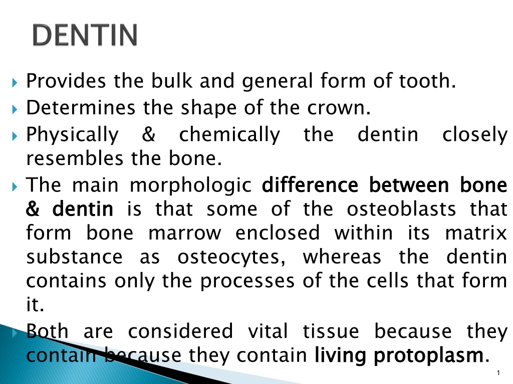
Provides the bulk and general form of tooth. Determines the shape of the crown. Physically resembles the bone. The main morphologic difference & & dentin form bone marrow enclosed within its matrix substance as osteocytes, whereas the dentin contains only the processes of the cells that form it. Both are considered vital tissue because they contain because they contain living & chemically the dentin closely difference between between bone bone dentin is that some of the osteoblasts that living protoplasm protoplasm. 1
It is light yellowish with age. yellowish in color, becoming darker It is elastic elastic and subject to slight deformation. Harder than bone but softer than enamel. Lower renders it more Lower content of mineral more radiolucent mineral salts in dentin radiolucent than enamel. 3
Consists inorganic The fibrils mucopolysaccharides aminoglycans) The hydroxyapetite Consists of inorganic material The organic fibrils mucopolysaccharides (proteoglycans aminoglycans). . The hydroxyapetite as of 35 material. . organic substance and 35% % organic organic matter matter and and water water & & 65 65% % substance consists and consists of ground (proteoglycans and of collagenous substance and glycos collagenous substance a a ground of of glycos inorganic inorganic component as in component in bone, consists consists of of bone, cementum cementum & & enamel enamel. Organic constituents can be removed from the mineral by incineration incineration or or organic organic chelation chelation. 4
The bodies layer their cytoplasmic processes are included in the tubules in the mineralized matrix. bodies of layer on the odontoblasts the pulpal of the pulpal surface odontoblasts are surface of are arranged of the arranged in dentin, and only in a a on the the dentin, Each the predentin & calcified dentin within one tubule. Each cell cell gives gives rise rise to to one one process process, which traverses Terminates in a branching network at the junction with enamel or cementum. Tubules therefore characteristic of it. Tubules are found throughout normal dentin & are 5
The course follows a gentle curve in the crown, less so in the root, where it resembles S S in in shape shape. Starting at right angles from the pulpal surface, the first convexity of this doubly curved course is directed toward the apex of the tooth. These dentinoenamel and dentinocementum junctions. tubules end perpendicular to the Near the root tip & along the incisal edges and cusps the tubules are almost straight. 6
The ratio between the outer dentin outer and and inner inner surfaces surfaces of of dentin is about 5 5: :1 1. . The ratio between the numbers area about 4:1. numbers of of tubules tubules per per unit unit area on the pulpal and outer surfaces of dentin is There are more tubules per unit area in the crown than in the root. The dentinal tubules have lateral branches throughout dentin, which are termed canaliculi microtubules. canaliculi or A few dentinoenamel termed few dentinal dentinoenamel junction termed enamel dentinal tubules junction into enamel spindles tubules extend into the spindles. . extend through the enamel through the enamel. . These the are These are 9
The dentin that immediately dentinal It is more highly mineralized than intertubular dentin. It is twice as thick in outer dentin (approx. 0.75um) than in inner dentin (0.4um). immediately surrounds surrounds the the dentinal tubules tubules. . 11
Forms the main body of dentin. main body of dentin. It is located between the dentinal tubules or, more specifically, between the zones of peritubular dentin. Its organic matrix is retained after decalcification. About one specifically collagen fibers. The fibrils range from 0.5 to 0.2um in diameter and exhibit crossbanding 64 one- -half half of of its its volume volume is is organic organic matrix matrix, crossbanding at for collagen at 64um um intervals, intervals, which which is is typical typical for collagen. . 14
Is located adjacent to the pulp tissue Is located adjacent to the pulp tissue. Is 2 2 to the odontoblast. to 6 6 um um wide wide, depending on the activity of It is the first formed dentin and is not mineralized. first formed dentin and is not mineralized. As the collagen fibers undergo mineralization at the predentin- dentin front, the predentin then becomes dentin and a new layer of predentin forms circumpulpally. 15
They are the cytoplasmic odontoblasts cytoplasmic extensions extensions of of the the odontoblasts. . The at processes The odontoblasts at the processes extend odontoblasts reside the pulp extend into reside in predentin border into the the peripheral border and the dentinal in the peripheral pulp and their dentinal tubules pulp their pulp- - predentin tubules. The processes are largest in diameter near the pulp and taper further into dentin. The odontoblast cell bodies are approximately 7 7um um in in diameter diameter and and 40 40um um in in length length. . 17
RELATIONSHIP BETWEEN ODONTOBLASTIC PROCESS AND DENTINAL TUBULE 18
Mantle underlying the dentinoenamel junction. It is the outer dentin & is about 20um thick. The fibrils found in this zone are perpendicular to the dentinoenamel junction. Mantle dentin dentin is the first formed dentin in the crown outer or or most most peripheral peripheral part part of the primary Circumpulpal dentin or bulk of the tooth. Represents all of the dentin completion The fibrils are much smaller in diameter & are more closely packed together. Slightly more mineral content than mantle dentin. Circumpulpal dentin dentin forms the remaining primary dentin formed formed prior prior to to root root completion. 19
A narrow band of dentin bordering the pulp and representing the dentin completion dentin formed formed after after root root completion. . Contains fewer tubules fewer tubules than primary dentin. There is usually a bend in the tubules where primary and secondary dentin interface. 21
The incremental lines They run at right angles to the dentinal tubules. These lines reflect the daily deposition in the daily formative process. The course of the lines indicates the growth pattern of the dentin. incremental lines lines, appear as fine lines or striations in dentin. lines (von (von ebner), ebner), or or imbrication imbrication daily rhythmic, rhythmic, recurrent recurrent deposition of dentin matrix as well as hesitation Some of the incremental lines are accentuated because of disturbances mineralization contour disturbances in process. . Such lines are known as lines of in the the matrix matrix and and mineralization process contour lines of owen owen. . 24
These These lines lines represent represent hypocalcified hypocalcified bands bands. . In the deciduous teeth and in the first permanent molars, the prenatal and postnatal dentin is separated by an accentuated contour line. This is termed the the neonatal neonatal line line. . This environment line reflects the abrupt abrupt change change in in environment that occurs at birth. The dentin matrix formed prior to birth is usually of better quality than that formed after birth. 27
Sometimes mineralization of dentin begins in small globular areas that fail to fuse into a homogenous mass. This results in zones of hypomineralization between interglobular dentin. hypomineralization between the the globules globules. . These zones are called Forms in crowns of teeth in the circumpulpal just circumpulpal dentin dentin just below below the the mantle mantle dentin dentin. . Follows an incremental pattern. The dentinal tubules pass uninterruptedly, thus demonstrating a defect matrix formation. defect of of mineralization mineralization & not of 29
There is a zone adjacent appears granular. This is known as Tomes granular adjacent to to the the cementum cementum that Tomes granular layer layer. . Caused by coalescing portions coalescing and of the and looping tubules. . looping of of the the terminal terminal portions of the dentinal dentinal tubules 32
Dentin Cementum Granular layer of Tomes Hyaline layer
Direct effect the nerve endings in the tubules. Direct conduction conduction theory theory in which stimuli directly Transduction odontoblast process conducts an impulse to the nerve endings in the predentin, odontoblast zone, and pulp. Transduction theory theory in which the membrane of the Fluid cause an inward or outward movement of fluid in the tubule, which in turn produces movement of the odontoblast and its processes. Fluid or or hydrodynamic hydrodynamic theory theory in which stimuli 35
The odontoblast processes disintegrate, & the empty tubules empty tubules are are filled filled with with air air. . Appear black in transmitted light & white in reflected light. Often observed in the area of narrow because of crowding of odontoblasts. narrow pulpal pulpal horns horns Demonstrate decreased sensitivity. Appear to a greater extent in older teeth. Probably the initial step in the formation of sclerotic dentin. 37
Dead tracts Tertiary dentin Secondary physiological dentin Primary physiological dentin
Collagen fibers and apatite crystals begin appearing in the dentinal tubules. Apatite crystals are initially only sporadic in a dentinal tubule but gradually fill it with a fine meshwork of crystals. Gradually, the tubule mineral, peritubular dentin. The refractive tubules are occluded are areas become transparent. Found specially in roots. Transparent or light reflected tubule lumen lumen is is obliterated obliterated with with mineral, which appears very much like the refractive indices indices of dentin in which the are equalized equalized, and such roots. light in in transmitted transmitted and dark dark in in reflected light light. . 39
Deantal caries Sclerotic dentin
Odontoblast layer Predentin Dentinal tubules