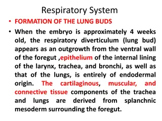
Understanding the Anatomy of Lungs
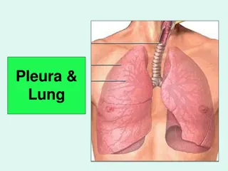
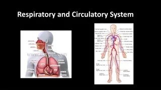
The lungs, vital organs of the respiratory system, consist of two cone-shaped structures with apex, base, costal, and medial surfaces. Positioned in the thoracic cavity, they are associated with the heart, great vessels, and other structures in the mediastinum. The right lung has three lobes, while the left lung has two due to the presence of the heart. The pleura and pleural cavity play a crucial role in lung function, with visceral and parietal pleura layers.

Download Presentation

Please find below an Image/Link to download the presentation.
The content on the website is provided AS IS for your information and personal use only. It may not be sold, licensed, or shared on other websites without obtaining consent from the author. Download presentation by click this link. If you encounter any issues during the download, it is possible that the publisher has removed the file from their server.
E N D
Presentation Transcript
LUNGS Done by: Mrs. Mercy Deva Priya Asst.Professor .Dept of MHN
Position and associated structures There are two lungs, one lying on each side of the midline in the thoracic cavity. They are cone-shaped and are described as having an apex, a base, costal surface and medial surface. The apex. This is rounded and rises into the root of the neck, about 25 mm (1 inch) above the level of the middle third of the clavicle. The structures associated with it are the first rib and the blood vessels and nerves in the root of the neck.
The base. This is concave and semilunar in shape and is closely associated with the thoracic surface of the diaphragm. The costal surface. This surface is convex and is closely associated with the costal cartilages, the ribs and the intercostal muscles. The medial surface. This surface is concave and has a roughly triangular-shaped area, called the hilum, at the level of the 5th, 6th and 7th thoracic vertebrae.
The area between the lungs is the mediastinum. It is occupied by the heart, great vessels, trachea, right andmleft bronchi, oesophagus, lymph nodes, lymph vessels and nerves.
Organisation of the lungs The right lung is divided into three distinct lobes: superior, middle and inferior. The left lung is smaller as the heart is situated left of the midline. It is divided into only two lobes: superior and inferior.
Pleura and pleural cavity The pleura consists of a closed sac of serous membrane (one for each lung) which contains a small amount of serous fluid. The lung is invaginated into this sac so that it forms two layers: one adheres to the lung and the other to the wall of the thoracic cavity The visceral pleura. The parietal pleura.
The visceral pleura. This is adherent to the lung, covering each lobe and passing into the fissures which separate them. The parietal pleura. This is adherent to the inside of the chest wall and the thoracic surface of the diaphragm. It remains detached from the adjacent structures in the mediastinum and is continuous with the visceral pleura round the edges of the hilum.
The pleural cavity. This is only a potential space. In health, the two layers of pleura are separated by only a thin film of serous fluid which allows them to glide over each other, preventing friction between them during breathing. The serous fluid is secreted by the epithelial cells of the membrane.
Interior of the lungs The lungs are composed of the bronchi and smaller air passages, alveoli, connective tissue, blood vessels, lymph vessels and nerves. The left lung is divided into two lobes and the right, into three Each lobe is made up of a large number of lobules.
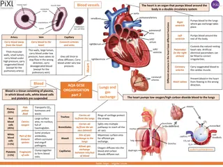
Pulmonary blood supply The pulmonary artery divides into two, one branch conveying deoxygenated blood to each lung. Within the lungs each pulmonary artery divides into many branches which eventually end in a dense capillary network around the walls of the alveoli. The exchange of gases between air in the alveoli and blood in the capillaries takes place across these two very fine membranes.
The pulmonary capillaries join up, becoming two pulmonary veins in each lung. They leave the lungs at the hilum and convey oxygenated blood to the left atrium of the heart. The innumerable blood capillaries and blood vessels in the lungs are supported by connective tissue.