Understanding Pleura and Lung Anatomy
Pleura is a double-layered serous membrane surrounding the lungs, consisting of parietal and visceral layers. The pleural cavity between them contains pleural fluid. Parietal pleura is subdivided into cervical, costal, mediastinal, and diaphragmatic regions. The pleura has nerve supply that makes it sensitive to pain, pressure, and touch. Understanding the anatomy of pleura and lungs is essential for knowledge of lung structures, relations, nerve supply, and blood supply.

Download Presentation

Please find below an Image/Link to download the presentation.
The content on the website is provided AS IS for your information and personal use only. It may not be sold, licensed, or shared on other websites without obtaining consent from the author. Download presentation by click this link. If you encounter any issues during the download, it is possible that the publisher has removed the file from their server.
E N D
Presentation Transcript
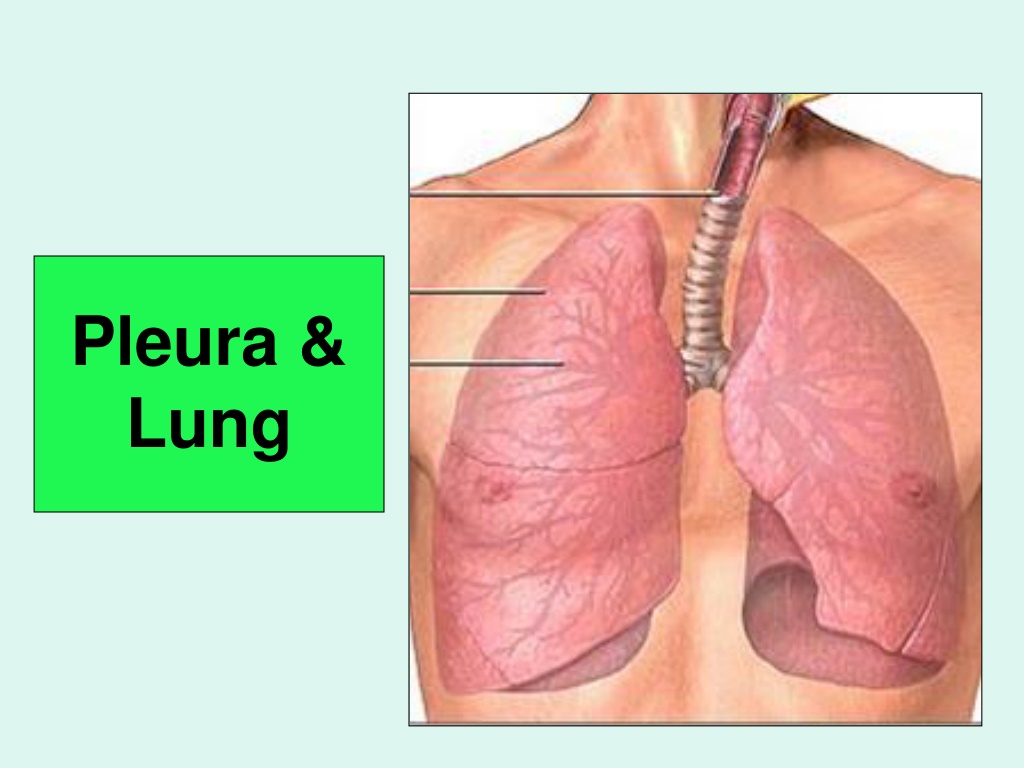
Pleura & Lung
Objectives By the end of the lecture, the student should be able to : Describe the anatomy of the pleura: subdivisions into parietal & visceral pleurae, nerve supply of each of them. List the parts of parietal pleura and its recesses. Describe the surface anatomy of both pleurae and lungs. Describe the anatomy of lungs : shape, relations, nerve supply & blood supply. Describe the difference between right & left lungs. Describe the formation of bronchopulmonary segments and the main characteristics of each segment in the lung.
Pleura Double-layered serous membrane enclosing the lung. Has two layers: Parietal layer, which lines the thoracic walls. Visceral layer, which covers the surfaces of the lung. The two layers continue with each other around the root of the lung, where it forms a loose cuff hanging down called the pulmonary ligament. The space between the two layers, the pleural cavity, contains a thin film of pleural serous fluid ( 5-10 ml.). Pulmonary ligament
Parietal Pleura It is divided according to the region in which it lies and the surfaces it covers, into: 1- Cervical 2- Costal 3- Mediastinal 4- Diaphragmatic Cervical
Parietal Pleura Cervical Pleura: Projects up into the neck about one inch above the medial1/3rdof clavicle. It lines the under surface of the suprapleural membrane. Costal pleura: lines, the back of the: Sternum, Ribs & costal cartilages, Intercostal spaces & Sides of vertebral bodies
Parietal Pleura Mediastinal pleura: covers the mediastinum. At the hilum, it is reflected on to the vessels and bronchi, and continuous with the visceral pleura. Diaphragmatic pleura: covers the thoracic (upper) surface of the diaphragm.
Pleural Recesses Costodiaphragmatic: Slit like space between costal and diaphragmatic pleurae, along the inferior border of the lung which enters through it in deep inspiration. Costomediastinal: Slit like space between costal and mediastinal pleurae, along the anterior border of the lung which enters through it in deep inspiration.
Pleura: Nerve Supply Parietal pleura: It is sensitive to pain, pressure, temperature, and touch. It is supplied as follows: Costal pleura is segmentally supplied by the intercostal nerves. Mediastinal pleura is supplied by phrenic nerves. Diaphragmatic pleura is supplied over the domes by phrenic nerves, around the periphery by lower 6 intercostal nerves. Visceral pleura sensitive to stretch only and is supplied by the autonomic fibers from the pulmonary plexus.
SURFACE ANATOMY OF PLEURA Apex: lies one inch above the medial 1/3 of the clavicle. The anterior margin Right pleura: extends vertically from sterno-clavicular joint to xiphisternal joint (6thcostal cartilage). Left pleura: Simillar course but at the level the 4thcostal cartilage deviates laterally and extends to lateral margin of the sternum to form cardiac notch then turns sharply downward to xiphisternal joint ( 6thcostal cartilage). Inferior margin : passes around the chest wall, on the 8thrib in midclavicular line, 10th rib in mid-axillary line and finally reaching to 12thrib adjacent to vertebral column posteriorly (T12 spine). Posterior margin : along the vertebral column from the apex (C7) to the inferior margin ( T12 spine). 4 6 6
SURFACE ANATOMY OF LUNG Apex, anterior border correspond nearly to the lines of pleura but are slightly away from the median plane. Inferior margin : passes around the chest wall, on the 6thrib in midclavicular line, 8th rib in mid-axillary line and finally reaching to 10thrib adjacent to vertebral column posteriorly. as the pleura but more horizontally and finally reaching to the 10ththoracic spine. Posterior margin : along the vertebral column from the apex (C7) to the inferior margin ( T10 spine).
SURFACE ANATOMY OF LUNG Oblique fissure: Represented by a line extending from 4th thoracic spine, obliquely ending at 6thcostal cartilage. Transverse fissure: Only in the right lung: represented by a line extending from 4thright costal cartilage to meet the oblique fissure.
Pleural Effusion It is an abnormal accumulation of pleural fluid about 300 ml, in the Costodiaphragmatic pleural recess , (normally 5-10 ml fluid) Causes: inflammation, TB, congestive heart disease and malignancy. The lung is compressed & the bronchi are narrowed. Auscultation would reveal only faint & decreased breathing sounds over compressed or collapsed lung lobe. Dullness on percussion over the effusion.
Lungs Located in the thoracic cavity, one on each side of the mediastinum Each lung is: Conical in shape. Covered by the visceral pleura. Suspended free in its own pleural cavity. Attached to the mediastinum only by its root.
LUNGS Each lung has: Apex and base: identify the top and bottom of the lung, respectively. Costal (lateral )surface: surrounded by the ribs from front & back). Medial (mediastinal) surface: Where the bronchi, blood vessels, and lymphatic vessels enter the lung at the hilum. It is also related to the structures forming the mediastinum.
LUNGS Apex: Projects into the root of the neck (1 inch above medial 1/3 of clavicle). It is covered by cervical pleura. It is grooved anteriorly by subclavian artery. Base: inferior or diaphragmatic surface) is concave and rests on the diaphragm.
Borders: Anterior & Posterior Anterior border : Is sharp, thin and overlaps the heart. Anterior border of left lung presents a cardiac notch at its lower end, has a thin projection called the lingula below the cardiac notch. Posterior border : is rounded, thick and lies beside the vertebral column.
Surfaces: Costal & Mediastinal Costal surface: Convex. Covered by costal pleura which separates lung from: ribs, costal cartilages & intercostal muscles. Medial surface: It is divided into 2 parts: Anterior (mediastinal) part: Contains a hilum in the middle (it is a depression in which bronchi, vessels, & nerves forming the root of lung). Posterior (vertebral) part: It is related to: Bodies of thoracic vertebrae, Intervertebral discs, Posterior intercostal vessels Sympathetic trunk. Lateral (costal) & medial surfaces of right lung
RIGHT LUNG ROOT 2 bronchi: Lie posterior. Pulmonary artery: Is superior Pulmonary veins: Are inferior and anterior.
LEFT LUNG ROOT One bronchus: Lies posterior Pulmonary artery: Is superior Pulmonary veins: Is inferior and anterior
Right lung Larger & shorter than left lung. Divided by 2 fissures (oblique & horisontal) into 3 lobes (upper, middle and lower lobes).
Left Lung Divided by one oblique fissure into -2 lobes, Upper and lower. There is No horizontal fissure. It has a cardiac notch at lower part of its anterior border.
Mediastinal surface of right lung On the mediastinal surface of the right lung, you find these structures: Azygos vein and its arch (posterior and over the root of the lung). Vagus nerve posterior to the root of the lung. Phrenic nerve anterior to the root of the lung. Cardiac impression: related to right atrium. Esophagus posterior to the root. Below hilum and in front of pulmonary ligament : groove for I.V.C. Cardiac impression
Mediastinal surface of left lung On the mediastinal surface of the left lung, you will find these structures: Descending aorta and its arches posterior and over to the root of the lung).. Vagus nerve posterior to the root of the lung over the root of the lung Phrenic nerve anterior to the root of the lung. Cardiac impression: related to left ventricle. Groove for left common carotid and left subclavian arteries Cardiac impression
Blood supply of lung Bronchial arteries (From descending aorta) . It supplies oxygenated blood to bronchi , lung tissue & visceral pleura. Bronchial veins : drain into azygos & hemiazygos veins. Pulmonary artery which carries non-oxygenated blood from right ventricle to the lung alveoli. 2 pulmonary veins : carry oxygenated blood from lung alveoli to the left atrium of the heart.
Nerve Supply of the lung Pulmonary plexus at the root of lung .is formed of autonomic N.S. from sympathetic & parasympathetic fibers. 1- Sympathetic Fibers From sympathetic trunk Action: broncho-dilatation/and vasoconstriction. 2- Parasympathetic Fibers From ..Vagus nerve . Action: broncho-constriction and vasodilatation and secretomotor to bronchial glands.
Bronchi The trachea divides into 2 main bronchi: Right main bronchus: which divides before entering the hilum, it gives: superior lobar (secondary) bronchus. On entering hilum, it divides into middle & inferior lobar bronchi. Left main bronchus: On entering hilum, it divides into superior & inferior lobar bronchi.
Bronchopulmonary segments They are the anatomic, functional, and surgical units of the lungs. Each lobar (secondary) bronchus gives segmental (tertiary) bronchi. Each segmental bronchus divides repeatedly into bronchioles. Bronchioles divide into terminal bronchioles, which show delicate outpouchings the respiratory bronchioles .
Bronchopulmonary segments The respiratory bronchioles end by branching into alveolar ducts, which lead into alveolar sacs. The alveolar sacs consist of several alveoli, each alveolus is surrounded by a network of blood capillaries for gas exchange. Alveolar sacs
Bronchopulmonary segments The main characteristics of a bronchopulmonary segment/ It is a subdivision of a lung lobe. It is pyramidal shaped, its apex toward the lung root. It is surrounded by connective tissue septa. It has a segmental bronchus, a segmental artery, lymph vessels, and autonomic nerves. The segmental vein lies in the inter- segmental C.T. septa between the segments. A diseased segment can be removed surgically, because it is a structural unit.