Exploring Microscopic Life Through a Microscope
Journey into the microscopic world by examining Elodea leaves and Paramecium under a microscope. Discover the intricate details of these tiny organisms, observe their movement, and learn about their structures. Engage in hands-on activities to explore the fascinating realm of microscopic life.

Download Presentation

Please find below an Image/Link to download the presentation.
The content on the website is provided AS IS for your information and personal use only. It may not be sold, licensed, or shared on other websites without obtaining consent from the author. Download presentation by click this link. If you encounter any issues during the download, it is possible that the publisher has removed the file from their server.
E N D
Presentation Transcript
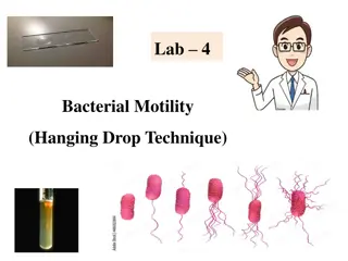
Day 1- Elodea Leaves Last week we looked at our cheek cells under the microscope Today, we are going to be looking at a different kind of microscopic life Open your folders to page 15
Page 15 Start with 100x magnification Describe what you see Increase to 400x magnification Describe what you see and draw it in the field of view Estimate the size of the cells in the leaf Record any additional observations
Slide set up Get a slide and a dropper from your microscope kit. Bring it to the Elodea station Break off half a leaf from the stem Place the leaf, top side up, on your slide Using your dropper, add a drop of the Elodea water to your slide Cover with a coverslip
Discussion What do you see when you look at the Elodea? Do the cells look empty? How many layers of cells do you see? 2, one large and one small
Discussion Continued Are all of the Elodea cells the same size? No, some are big and some are small Are the cells on the large cells on the top of the leaf or bottom of the leaf? The larger cells are on top How big are the Elodea cells? The larger ones are about 0.1 mm and the smaller ones are about 0.05 mm
Day 2- Paramecium Slide set up Use a dropper to collect some water from the bottom of the paramecium container Add 1 drop to your slide Place a VERY SMALL amount of spread out cotton on top Cover with a cover slip
Finding the Paramecium Start on 40x and look for tiny ovals moving around Open to page 16 of your notebook and answer the questions After you find your paramecium, change the lighting using the diaphragm to see different details
Discussion What movements did you observe? The paramecium was moving around the slide and there were moving circles inside the paramecium What did you see on the inside of the paramecium? Blobs, circles, dark areas What did you see on the outside of the paramecium? Little legs or hairs How big was the paramecium? Less than 0.5 mm
Are they living or nonliving? living What is your evidence? They are moving Could you see it eat or use energy? Give off waste? Reproduce? no What might we do to see some of these activities? Feed them
Protists Paramecia are single-celled organisms in the Protista kingdom In Greek, proto means early and protist means the very first The single-celled organisms like paramecia are members of a kingdom of life that are similar to some of the very first life-forms on Earth. Protists are not animals, animals are always multicellular Protista is a separate kingdom whose members are mostly single-celled
Living vs. Organism Each Elodea cell is living while the paramecia cell is an organism. The Elodea cells stay in one place; paramecia move around Elodea cells are stuck together; paramecia cells are alone Elodea cells are part of a bigger organism; paramecia are not
Paramecia Feeding Prepare a wet mount of paramecia with cotton just like before. Add one drop of the Congo red-dyed yeast Add the cover slip Locate a paramecium and look for evidence that it is eating yeast Observe first at 100x and then at 400x. Record observations in words and illustration
Color Change Congo red does two things It makes the yeast easier to see It also acts as an acid indicator What happened when acid was added to the Congo red? How can Congo red be used as an indicator?
Things to think about Paramecia do not have legs or fins. How do you think they move around? How do the paramecia get the yeast inside their bodies? Do paramecia have mouths? How do paramecia get rid of waste? What keeps the paramecium s insides from spilling out? What does the yeast look like inside a paramecium?
Lets Talk What did you observe? If you noticed bubbles inside the paramecium getting bigger and then appearing to burst, those are called water-expelling vesicles Hairlike structures covering exterior of the paramecia are called cilia and help the paramecia move