Bacterial Motility Testing Using Hanging Drop Technique
The Hanging Drop Technique is a method used to determine the motility of bacteria by suspending them in a drop of fluid on a slide. This procedure involves preparing a hanging drop slide, inoculating the drop with bacteria, sealing it with petroleum jelly, and observing the organisms under a microscope. Motility testing is crucial in microbiology for identifying motile organisms like Proteus vulgaris and Staphylococcus epidermidis.

Download Presentation
Please find below an Image/Link to download the presentation.
The content on the website is provided AS IS for your information and personal use only. It may not be sold, licensed, or shared on other websites without obtaining consent from the author. Download presentation by click this link. If you encounter any issues during the download, it is possible that the publisher has removed the file from their server.
Presentation Transcript
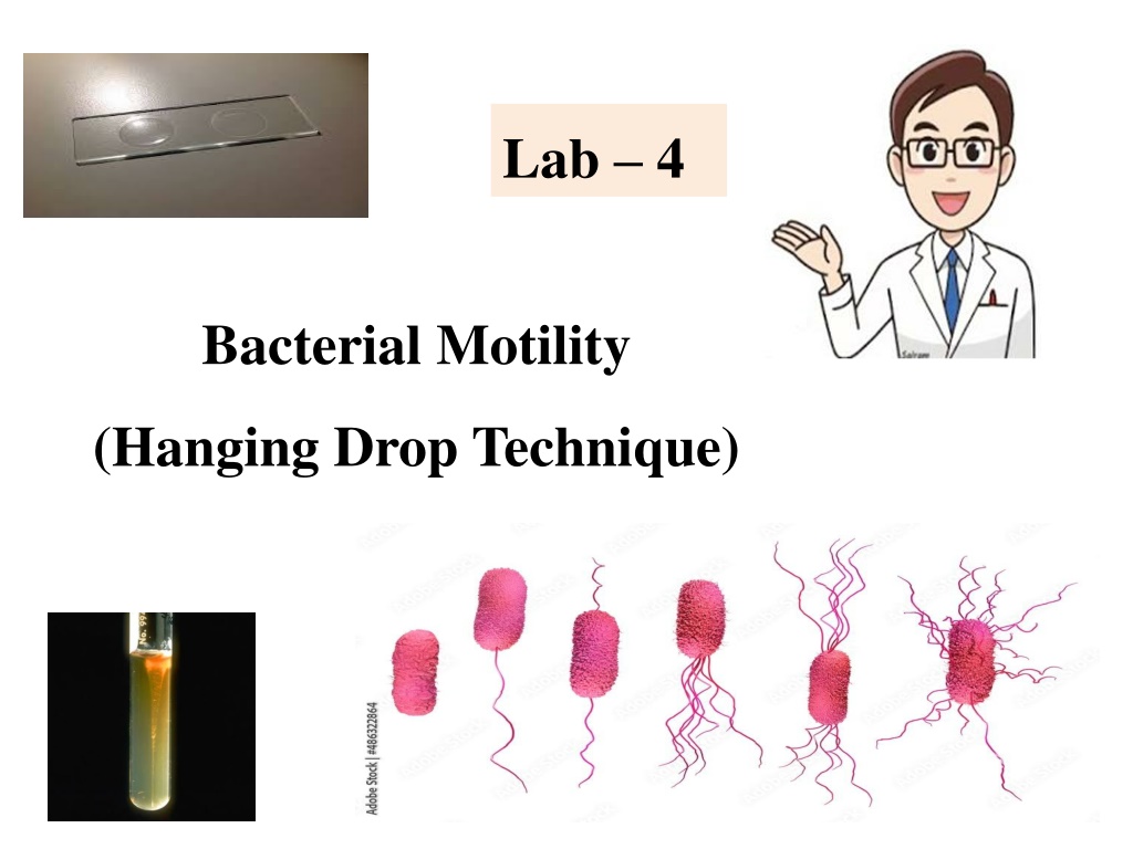
Lab 4 Bacterial Motility (Hanging Drop Technique)
Motility testing Motility could be detected by: 1) 2) 3) Hanging Drop technique. Semi-Solid media Inoculation. Flagella stain.
1. The Hanging-Drop Preparation; The simplest method for examining living microorganisms is to suspend them in a fluid (water, saline, or broth) and prepare a ''hanging drop,'' using a cover glass and a hollow ground slide. The slide is ground with a concave well in the center; the cover glass holds a drop of the suspension. When the cover glass is inverted over the well of the slide, the drop hangs from the glass in the hollow concavity of the slide. Microscopic study of such a wet preparation can provide very useful information. This method is used to determine whether an organism is motile or not. Such a slide can be observed for a fairly long time period, because the drop does not dry up quickly.
Hanging Drop slide The slide for a hanging drop is ground with a concave well in the centre; the cover glass holds a drop of the suspension. When the cover glass is inverted over the well of the slide, the drop hangs from the glass in the hollow concavity of the slide. Since the drop lies within an enclosed glass chamber, drying out occurs very slowly. A ring of Vaseline around the edge of the cover slip keeps the slide from drying out.
Objective : To observe bacteria in a wet mount and determine their motility. Materials 24-hour broth culture of Proteus vulgaris 24-hour broth culture of Staphylococcus epidermidis. 2 hollow-ground slide Several cover glasses Wire inoculating loop Bunsen burner China-marking pencil Petroleum jelly
Procedures 1. Take a cover glass and clean it thoroughly, making certain it is free of grease (the drop to be placed on it will not hang from a greasy surface). It may be dipped in alcohol and polished dry with tissue; or washed in soap and water, rinsed completely, and wiped dry. 2. Take one hollow-ground slide and clean the well with a piece of dry tissue. Place a film of petroleum jelly around the rim of the well. 3. Gently shake the broth culture of Proteus until it is evenly suspended. Using the wire inoculating loop, sterilize on the Bunsen flame, remove a loopful of culture. Close, and return the tube to the rack. 4. Place the loopful of culture in the center of the cover glass (do not spread it around). Flame the loop and put it down. 5. Hold the hollow-ground slide inverted, well down, over the cover glass; then press it down lightly so that the petroleum jelly adheres to the four edges of the cover glass. Now turn the slide over. You should have a sealed wet mount, the drop of culture hanging in the well.
6. Place the slide on the microscope stage, cover glass up. Start your examination with the low-power objective to find the focus. It is helpful to focus first on one edge of the drop, which will appear as a dark line. 7. The light should be reduced with the iris diaphragm, and, if necessary, by lowering the condenser. If you have trouble with the focus, ask the instructor for help. 8. Continue your examination with the high-dry and oil immersion objectives (be very careful not to break the cover slip with the latter). 9. Make a hanging-drop preparation of the staphylococcus culture, following the same procedure described above. 10. Record your observation of the shape, cell groupings, and motility of the organisms. 11. Discard your slides in a container with disinfectant solution.
cover glass Bacterial suspension petroleum Hollow ground slide concave well
The most commonly used test for motility in microbiology lab. It depends on the ability of motile bacteria to move through semi-solid media. Ordinary solid media contain 1.5-2.0% Agar Semi solid media contain about 0.5- 1% Agar
Procedure of Motility Test How to Perform Test: Using a sterile bacteriological needle, pick a colony of the test organism Stab quickly a tube of semi solid media. (avoid using bent needles). Incubate the semi solid media for 24 hours
3. Flagella staining 1. Rosanalin dye 2. Silver nitrate + ferric tannate
Site of flagella Lophotrichous (pseudomonas) (Spirillum volutans) amphitrichous
Site of flagella Peritrichous (E.coli) Monotrichous ( Vibrio cholerae)
Types of motility 1-True movement (depends on flagella) 2- False movement. a. Brownian movement (Vibratory movement) b. Drafting movement - organisms streaming along a tide