Microscope Focusing and Bacteria Morphology Guide
Learn how to focus a microscope for simple staining, understand the morphology of bacteria such as cocci, bacillus, spirals, and pleomorphic shapes, and discover the process of preparing a slide smear with detailed steps and images.

Download Presentation

Please find below an Image/Link to download the presentation.
The content on the website is provided AS IS for your information and personal use only. It may not be sold, licensed, or shared on other websites without obtaining consent from the author. Download presentation by click this link. If you encounter any issues during the download, it is possible that the publisher has removed the file from their server.
E N D
Presentation Transcript
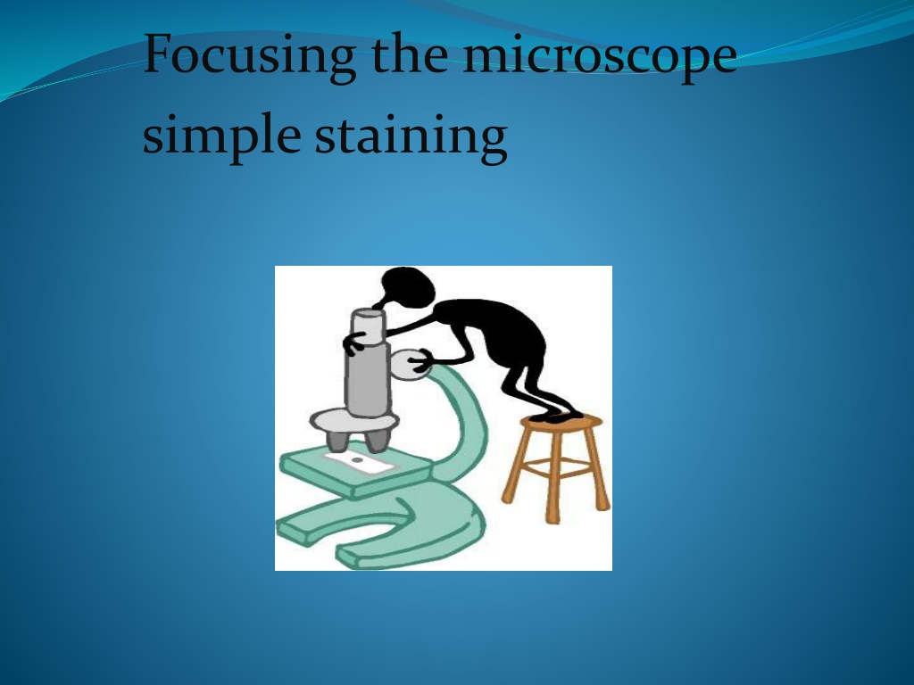
Focusing the microscope simple staining
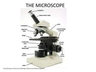
How to Focus the Microscope? Turn the revolving nosepiece to 10 magnifying lens. Move the stage up, the condenser to the top position and close iris diaphragm half way. 3. Put the slide on the slide holder on the stage. 4. Put the stained area (field) under the objective lens using the stage control knobs. Make eye adjustment. Focus using coarse adjustment knob then by fine adjustment knob. 1. 2. 5.
6. Move to Magnification 100 objective lens once you are in focus with x10 lens. Don't forget to put a drop of oil immersion on the slide. 7. Open iris diaphragm (more light). 8. Use fine adjustment knob to focus .Don't use coarse adjustment knob at this stage. 9. Look for proper area, when you can see the organism s shape clearly.
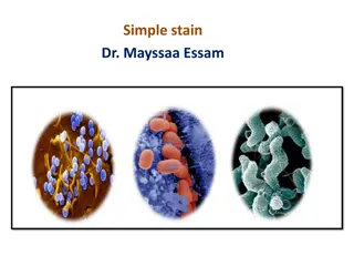
Morphology of the Bacteria: 1) Coccus (single), cocci (sphere). Arrangement: Single (round) Diplococci (pairs). Streptococci (chain). Staphylococci (group, like clusters). Micrococci (in packets of 4 or 8).
2) Bacillus (single), bacilli (group). Arrangement: Rod. Diplobacilli Streptobacilli (chain). Fusiform (pointed end). Coccobacilli.
3) Spiral Arrangement: Spirillum (one coil). Spirochetes (Many coils) Comma shape. 4) Pleomorphic has different shapes (Chinese letter).
Preparing a slide (smear) Open the Bunsen burner or Bacti-burner. Label the slide: organism s name, stain, your name. Sterilize the loop: heat the loop till it become red, then wait few seconds to cool (don t kill the organism) Deliver 1-2 drops of saline on the slide. Sterilize the loop. By a gentle touch take a colony of organism from culture by the loop.
Spread the organism on the slide in an oval shape. Sterilize the loop. Leave the smear at room temperature to air dry. Put the slide on the slide warmer(40 C) 5 min to: Fix the organism on slide: the organism will be fixed and stick on the slide by protein coagulation due to heating. 1. 2. Kill the organism.
Switch off the flame. Stain: Put the slide on the staining rack (slide upward) 1. 2. Cover the entire organism with the stain (methylene blue) on the slide for 1 min. 3. Rinse with water. 4. Dry with filter paper by gentle pressing. Focus under the microscope and report your observations( need to report shape and arrangement)
Stain dye: Stain (dye):I t is a salts in which one of the salt ions (-ve or +ve) is colored. Ex. : Methylene blue. Basic dye: if the color is in the +ve ion of the dye. Acidic dye: if the color is in the -ve ion of the dye. Basic dye: MBCl MB++Cl- There is a slight ( ve) charge on the cell wall of living cell, the basic dye (+vecharge) will be attracted to bacteria and stain it.


 Bacillus (single), bacilli (group).")
 Spiral")
 Spiral")
")