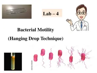
Microbiology Study: Hanging Drop Technique in Department of Microbiology, College of Medicine

In the Department of Microbiology at the College of Medicine, the Hanging Drop Technique is utilized to study live microorganisms. This technique involves suspending microorganisms in fluid on a hollow ground slide, allowing for observation of their morphology under a microscope. By creating a wet mount with a cover glass and a concave well slide, researchers can determine the motility of organisms, such as Proteus vulgaris and Staphylococcus epidermidis. The step-by-step procedures involve careful preparation and examination using different microscope objectives.

Download Presentation

Please find below an Image/Link to download the presentation.
The content on the website is provided AS IS for your information and personal use only. It may not be sold, licensed, or shared on other websites without obtaining consent from the author. Download presentation by click this link. If you encounter any issues during the download, it is possible that the publisher has removed the file from their server.
E N D
Presentation Transcript
Practical No. 4 Hanging Drop Technique Department of Microbiology College of Medicine
1. The Hanging-Drop Preparation; Since you have been oriented to some basic tools and methods used in microbiology, we shall begin our study of microorganisms by learning how to prepare them for study of their morphology under the microscope. The simplest method for examining living microorganisms is to suspend them in a fluid (water, saline, or broth) and prepare a ''hanging drop,'' using a cover glass and a hollow ground slide. The slide is ground with a concave well in the center; the cover glass holds a drop of the suspension. When the cover glass is inverted over the well of the slide, the drop hangs from the glass in the hollow concavity of the slide. Microscopic study of such a wet preparation can provide very useful information. Primarily the method is used to determine whether an organism is motile or not. Such a slide can be observed for a fairly long time period, because the drop does not dry up quickly.
Objective : To observe bacteria in a wet mount and determine their motility. Materials 24-hour broth culture of Proteus vulgaris 24-hour broth culture of Staphylococcus epidermidis. 2 hollow-ground slide Several cover glasses Wire inoculating loop Bunsen burner China-marking pencil Petroleum jelly
cover glass Bacterial suspension petroleum Hollow ground slide concave well
Procedures 1-Take a cover glass and clean it thoroughly, making certain it is free of grease (the drop to be placed on it will not hang from a greasy surface). It may be dipped in alcohol and polished dry with tissue; or washed in soap and water, rinsed completely, and wiped dry. 2-Take one hollow-ground slide and clean the well with a piece of dry tissue. Place a film of petroleum jelly around the rim of the well. 3- Gently shake the broth culture of Proteus until it is evenly suspended. Using the wire inoculating loop, sterilize on the Bunsen flame, remove a loopful of culture. Close, and return the tube to the rack. 4- Place the loopful of culture in the center of the cover glass (do not spread it around). Flame the loop and put it down. 5- Hold the hollow-ground slide inverted, well down, over the cover glass; then press it down lightly so that the petroleum jelly adheres to the four edges of the cover glass. Now turn the slide over. You should have a sealed wet mount, the drop of culture hanging in the well.
6- Place the slide on the microscope stage, cover glass up. Start your examination with the low-power objective to find the focus. It is helpful to focus first on one edge of the drop, which will appear as a dark line. The light should be reduced with the iris diaphragm, and, if necessary, by lowering the condenser. If you have trouble with the focus, ask the instructor for help. 7- Continue your examination with the high-dry and oil immersion objectives (be very careful not to break the cover slip with the latter). 8- Make a hanging-drop preparation of the staphylococcus culture, following the same procedure described above. 9- Record your observation of the shape, cell groupings, and motility of the organisms. 10- Discard your slides in a container with disinfectant solution.
Note: True independent motility of bacteria depends on their possession of flagella. If so equipped, they can propel themselves with progressive, directional locomotion (often quit rapidly). This kind of active motion must be distinguished from the vibratory movement of organisms or other particles suspended in the fluid. The latter type of motion is caused by the continuous, rapid oscillation of molecules of the fluid. Small particles of any kind, including bacteria (whether motile or not), are constantly bombarded by the vibration of the fluid molecules, and so are bobbed up and down, back and forth. Such movement is irregular and non-directional and does not cause non motile organisms to change position with respect to other objects around them. One must be careful not to mistake movement caused by currents in a liquid for true motility. If a wet mount is not well sealed or contains bubbles, air currents set up reaction fluid currents, and one sees organisms streaming along on a tide.
Thank You Thank You